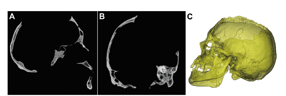
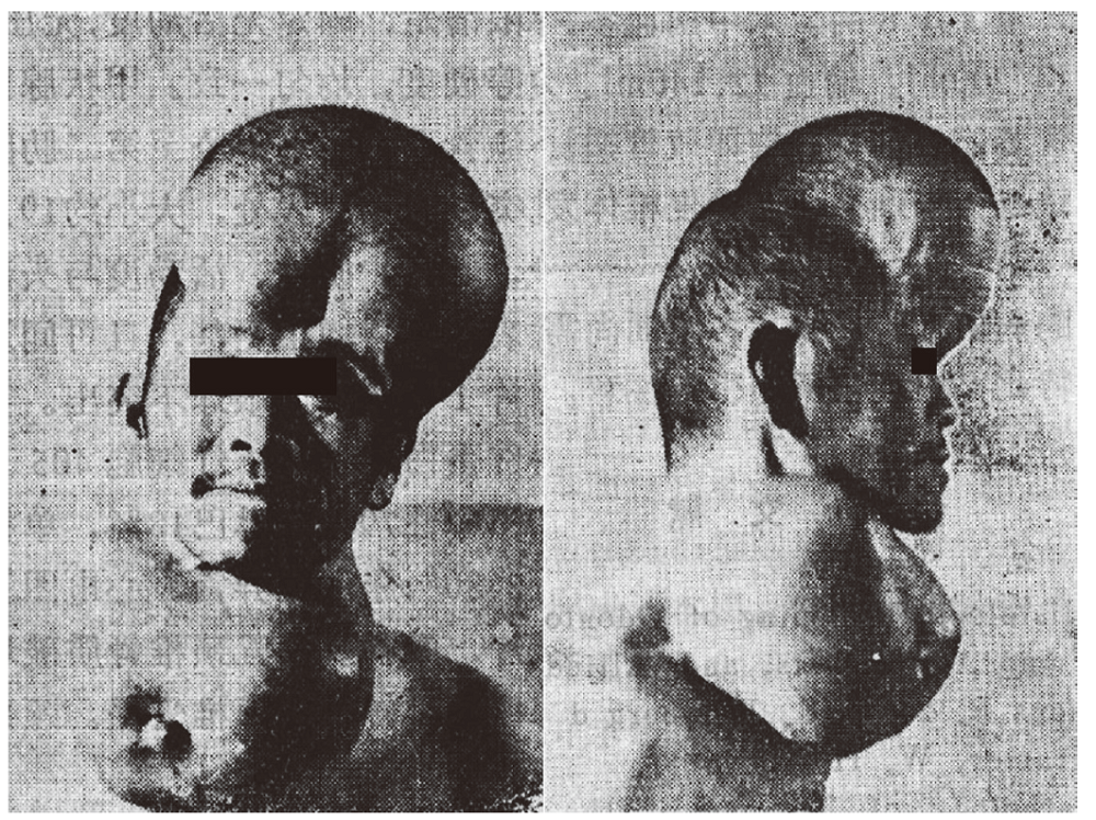
| [1] |
Marks MK, Hamilton MD. Metastatic Carcinoma: Palaeopathology and Differential Diagnosis[J]. International Journal of Osteoarchaeology, 2007,17:217-234
doi: 10.1002/(ISSN)1099-1212
URL
|
| [2] |
Binit S, Mittal MK, Mittal A, et al. Atypical lytic lesions of skull: Clinical and radiological correlation[J]. Annals of Indian Academy of Neurology, 2015,18:117-119
doi: 10.4103/0972-2327.144309
URL
pmid: 25745329
|
| [3] |
Lloret I, Server A, Taksdal I. Calvarial lesions: a radiological approach to diagnosis[J]. Acta Radiologica, 2009,50:531-542
doi: 10.1080/02841850902795274
URL
pmid: 19353343
|
| [4] |
Gomez CK, Schiffman SR, Bhatt AA. Radiological review of skull lesions[J]. Insights Imaging, 2018,9:857-882
doi: 10.1007/s13244-018-0643-0
URL
pmid: 30232767
|
| [5] |
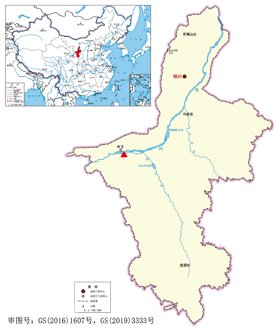
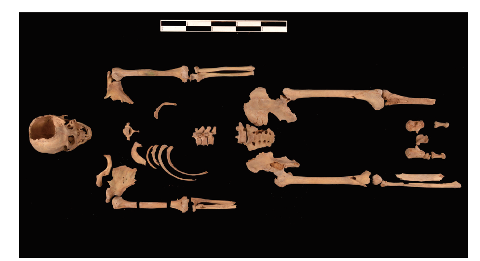
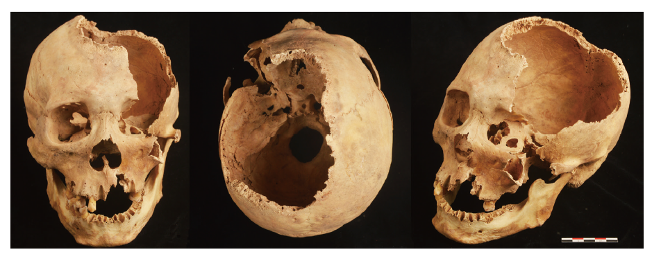
周赟, 王晓阳, 马强, 等. 宁夏海原石砚子汉墓发掘简报[J]. 文博, 2018,4:3-16
|
| [6] |
朱泓. 体质人类学[M]. 北京: 高等教育出版社, 2004: 92-106
|
| [7] |
邵象清. 人体测量手册[M]. 上海: 上海辞书出版社, 1985: 34-56
|
| [8] |
Buikstra JE, Ubelaker DH. Standards for data collection from human skeletal remains[M]. Fayetteville: Arkansas Archeological Survey, 1994: 15-38
|
| [9] |
Brooks S, Suchey JM. Skeletal age determination based on the Os pubis: Acomparison of the Acsádi-Nemeskéri and Suchey-Brooks methods[J]. Human Evolution, 1990,5:227-238
doi: 10.1007/BF02437238
URL
|
| [10] |
Colas L, Caron S, Cotton A. Skull vault lesions: a review[J]. American Journal of Roentgenology, 2015,205:840-847
|
| [11] |
Garfinkle J, Melançon D, Cortes M, et al. Imaging pattern of calvarial lesions in adults[J]. Skeletal Radiology, 2011,40(10):1261-1273
doi: 10.1007/s00256-010-0971-8
URL
pmid: 20526773
|
| [12] |
Yalçin Ö, Yildirim T, Kizilkiliç O, et al. CT and MRI findings in calvarial non-infectious lesions[J]. Diagnostic and Interventional Radiology, 2007,13:68-74
URL
pmid: 17562510
|
| [13] |
Marques C, Santos AL, Cunha E. Better a broader diagnosis than a misdiagnosis: the study of a neoplastic condition in a male individual who died in early 20th century (Coimbra, Portugal)[J]. International Journal of Osteoarchaeology, 2013,23(6):664-675
|
| [14] |
Berrettoni BA, Carter JR. Mechanisms of cancer metastasis to bone[J]. The Journal of bone and joint surgery, 1986,68(2):308-312
URL
pmid: 3753707
|
| [15] |
胥少汀, 葛宝丰, 徐印坎. 实用骨科学[M]. 北京: 人民军医出版社, 2015: 1777-1784
|
| [16] |
Marcove RC, Arlen M. Atlas of Bone Pathology: with Clinical and Radiographic Correlations[M]. Philadelphia: J.B. Lippincott, 1992
|
| [17] |
Body JJ. Metastatic bone disease[A]. In: Dynamics of bone and cartilage metabolism[C], MJ Seibel, SP Robins, JP Bilezikian (eds). San Diego: Academic Press, 1999: 793-810
|
| [18] |
Jaffe HL. Tumors and Tumorous Conditions of the Bones and Joints[M]. Philadelphia: Lea and Febiger, 1958
|
| [19] |
Dorfman HD, Czerniak B. Bone Tumors[M]. St. Louis: Mosby, 1998
|
| [20] |
Roodman GD. Mechanisms of bone metastasis[J]. The New England Journal of Medicine, 2004,350:1655-1664
doi: 10.1056/NEJMra030831
URL
pmid: 15084698
|
| [21] |
Ortner DJ, Putschar WGJ. Identification of Pathological Conditions in Human Skeletal Remains[M]. Washington, DC: Smithsonian Institution Press, 1981
|
| [22] |
Chew FS. Skeletal Radiology: The Bare Bones[M]. Maryland: Aspen Publishers, 1989
|
| [23] |
Strouhal E. Myeloma multiplex versus osteolytic metastatic carcinoma: differential diagnosis in dry bones[J]. International Journal of Osteoarchaeology, 1991,1:219-224
|
| [24] |
Waldron T. Lytic lesions in a skull: a problem of diagnosis[J]. Journal of Paleopathology, 1987,1:5-14
|
| [25] |
Khung S, Budzik JF, Amzallag-Bellenger E, et al. Skeletal involvement in Langerhans cell histiocytosis[J]. Insights Imaging, 2013,4(5):569-579
URL
pmid: 23907805
|
| [26] |
Hoover KB, Rosenthal DI, Mankin H. Langerhans cell histiocytosis[J]. Skeletal Radiology, 2007,36:95-104
URL
pmid: 17028900
|
| [27] |
Arana E, Martí-Bonmati L. CT and MR imaging of focal calvarial lesions[J]. American Journal of Roentgenology, 1999,172(6):1683-1688
doi: 10.2214/ajr.172.6.10350315
URL
pmid: 10350315
|
| [28] |
杨艳萍. 骨血管瘤CT和磁共振诊断及鉴别诊断[J]. 山西医药杂志, 2015,44(17):2000-2002
|
| [29] |
孙帮利, 刘晓红, 齐先龙. 骨血管瘤的影像学诊断[J]. 济宁医学院学报, 2002,25(2):38-39
|
| [30] |
Bastug D, Ortiz O, Schochet SS. Hemangiomas in the calvaria: imaging findings[J]. American Journal of Roentgenology, 1995,164(3):683-687
doi: 10.2214/ajr.164.3.7863894
URL
pmid: 7863894
|
| [31] |
贾雄, 刘晓红. 骨血管瘤的影像学诊断与病理分析研究[J]. 西北国防医学杂志, 2017,38(6):377-380
|
| [32] |
Jackson JBS. A boneless arm[J]. The Boston Medical and Surgical Journal, 1838,18:368-369
|
| [33] |
Gorham LW, Stout AP. Massive osteolysis (acute spontaneous absorption of bone, phantom bone, disappearing bone): its relation to hemangiomatosis[J]. The Journal of Bone & Joint Surgery, 1955,37(5):985-1004
URL
pmid: 13263344
|
| [34] |
Dellinger MT, Garg N, Olsen BR. Viewpoints on vessels and vanishing bones in Gorham-Stout disease[J]. Bone, 2014,63:47-52
URL
pmid: 24583233
|
| [35] |
Ruggieri P, Montalti M, Angelini A, et al. Gorham-Stout disease: the experience of the Rizzoli Institute and review of the literature[J]. Skeletal Radiology, 2011,40:1391-1397
doi: 10.1007/s00256-010-1051-9
URL
pmid: 20972870
|
| [36] |
赵书一, 刘天婧, 王恩波. Gorham-Stout综合征的研究进展[J]. 中华小儿外科杂志, 2020,3:280-284
|
| [37] |
黄曌殊, 范光明. 颅骨Gorham-Stout综合征一例[J]. 中国医学影像技术, 2015,31(11):1679
|
| [38] |
胥德政. Gorham综合征概述[J]. 中国疗养医学, 2011,20(2):146-148
|
| [39] |
李小虎, 王万勤, 陈华平, 等. 大块骨质溶解症的影像学表现(附2例报告及文献复习)[J]. 临床放射学杂志, 2010,29(11):1567-1569
|
| [40] |
Mirra JM, Picci RP, Gold RH. Bone Tumor: Clinical, Radiologic, and Pathologic Correlations[M]. London: Lea and Febiger, 1989
|
| [41] |
Aufderheide AC, Rodríguez-Martín C. The Cambridge Encyclopedia of Human Paleopathology[M]. Cambridge: Cambridge University Press, 1998: 351-354
|
| [42] |
Ortner DJ. Identification of Pathological Conditions in Human Skeletal Remains[M]. San Diego: Academic Press, 2003: 376-382
|
| [43] |
Roberts CA, Manchester K. The Archaeology of Disease[M]. Ithaca: Cornell University Press, 1995: 258-260
|
| [44] |
Fleming H. Osteomyelitis of the skull[J]. Cal West Med, 1925,23(8):985-988
URL
pmid: 18739729
|
| [45] |
Peterson L. Contemporary Oral Maxillofacial Surgery (4th ed)[M]. Philadelphia: Mosby Publishers, 2003: 375-377
|
| [46] |
Prasad KC, Prasad SC, Mouli N, et al. Osteomyelitis in the head and neck[J]. Acta Otolaryngol, 2007,127(2):194-205
doi: 10.1080/00016480600818054
URL
pmid: 17364352
|
| [47] |
Pincus DJ, Armstrong MB, Thaller SR. Osteomyelitis of the craniofacial skeleton[J]. Semin Plast Surg, 2009,23(2):73-79
doi: 10.1055/s-0029-1214159
URL
pmid: 20567729
|
| [48] |
李长山. 颅脑损伤并发症的 CT 与MRI 表现[J]. 医学影像学杂志, 2007,17(2):163
|
| [49] |
周应平, 程晓光, 刘桐希, 等. 骨嗜酸性肉芽肿与骨髓炎、尤文肉瘤的影像学比较[J]. 中国医药科学, 2013,3(7):119-122, 126
|
| [50] |
Kaul R, Gupta N, Gupta S, et al. Eosinophilic granuloma of skull bone[J]. Journal of Cytology, 2009,26(4):156-157
URL
pmid: 21938183
|
| [51] |
Capasso LL. Antiquity of cancer[J]. International Journal of Cancer, 2005,113(1):2-13
doi: 10.1002/ijc.20610
URL
pmid: 15389511
|
| [52] |
Anderson T, Wakely J, Carter AR. Medieval example of metastatic carcinoma: a dry bone, radiological, and SEM study[J]. American Journal of Physical Anthropology, 1992,89(3):309-323
URL
pmid: 1485639
|
| [53] |
De la Rúa C, Baraybar JP, Etxeberia F. Neolithic case of metastatic carcinoma: Multiple approaches to differential diagnosis[J]. International Journal of Osteoarchaeology, 1995,5(3):254-264
|
| [54] |
Grupe G. Metastazing carcinoma in a medieval skeleton: Differential diagnosis and etiology[J]. American Journal of Physical Anthropology, 1988,75(3):369-374
doi: 10.1002/ajpa.1330750308
URL
pmid: 3284377
|
| [55] |
Schultz M. A case of metastatic carcinoma from Christian Sayala (Egyptian Nubia)[J]. Anthropologisher Anzeiger, 1993,51(2):97-115
|
| [56] |
Gladykowska-Rzeczycka J. Tumors in antiquity in East and Middle Europe[A]. In: Ortner DJ, Aufderheide AC eds. Human Paleopathology-Current Syntheses and Future Options[C]. Washington: Smithsonian Institution Press, 1991: 251-256
|
| [57] |
Caruso V, Gibelli D, Castoldi E, et al. Metastatic Cancer in the Middle Age: The Possible Case of a Female Skeleton from Bormio (Italy)[J]. International Journal of Osteoarchaeology, 2017,27(6):1022-1037
|
| [58] |
Manchester K. Secondary cancer in an Anglo-Saxon female[J]. Journal of Archaeological Science, 1983,10(5):475-482
|
| [59] |
Møller P, Møller-Christensen V. A medieval female skull showing evidence of metastases from a malignant growth[J]. Acta Pathologica et Microbiologica Scandinavica, 1952,30(3-4):336-342
doi: 10.1111/j.1699-0463.1952.tb00186.x
URL
pmid: 14933065
|
| [60] |
Ortner DJ, Manchester K, Lee F. Metastatic carcinoma in a leper skeleton from a medieval cemetery in Chichester, England[J]. International Journal of Osteoarchaeology, 1991,1(2):91-98
|
| [61] |
Šefčáková A, Strouhal E, Němečková A, et al. Case of metastatic carcinoma from end of the 8th-Early 9th Century Slovakia[J]. American Journal of Physical Anthropology, 2011,116(3):216-229
doi: 10.1002/ajpa.1117
URL
pmid: 11596001
|
| [62] |
Tkocz I, Bierring F. A medieval case of metastasizing carcinoma with multiple osteosclerotic bone lesions[J]. American Journal of Physical Anthropology, 1984,65(4):373-380
URL
pmid: 6395693
|
| [63] |
Wakely J, Anderson T, Carter A. A multidisciplinarian case study of prostatic(?) carcinoma from medieval Canterbury[J]. Journal of Archaeological Science, 1995,22(4):469-477
|
| [64] |
Schultz M, Parzinger H, Posdnjakov DV, et al. Oldest known case of metastasizing prostate carcinoma diagnosed in the skeleton of a 2,700-year-old Scythian King from Arzhan (Siberia, Russia)[J]. International Journal of Cancer, 2007,121(12):2591-2595
doi: 10.1002/ijc.23073
URL
pmid: 17918181
|
| [65] |
Wasterlain SN, Ascenso BF, Silva AM. Skeletal metastatic carcinoma: A case from 15th-20th century Coimbra, Portugal[J]. International Journal of Osteoarchaeology, 2011,21(3):336-346
|
| [66] |
Schats R, Hoogland M, Waters-Rist A. A probable case of metastatic carcinoma in the medieval Netherlands[J]. International Journal of Paleopatholoy, 2018,22:181-188
|
| [67] |
Horáčková L, Benešová L, Strouhal E, et al. A case of severe metastatic carcinoma in a Late Medieval skull from Petrov, Brno (Czech Republic)[J]. Anthropologie, 1997,35(1):57-64
|
| [68] |
Assis S, Codinha S. Metastatic carcinoma in a 14th-19th century skeleton from Constância (Portugal)[J]. International Journal of Osteoarchaeology, 2009,20(5):603-620
|
| [69] |
Melikian M. A case of metastatic carcinoma from 18th century London[J]. International Journal of Osteoarchaeology, 2006,16(2):138-144
|
| [70] |
Strouhal E, Vyhnanek L. New cases of malignant tumors from late period cemeteries at Abusir and Saqqara (Egypt)[J]. Ossa, 1982,8:165-189
|
| [71] |
Wells C. Ancient Egyptian Pathology[J]. Journal of Laryngology and Otology, 1963,77(3):261-265
|
| [72] |
Nerlich AG, Rohrbach H, Bachmeier B, et al. Malignant tumors in two ancient populations: An approach to historical tumor epidemiology[J]. Oncol Rep, 2006,16(1):197-202
URL
pmid: 16786146
|
| [73] |
Prates C, Sousa S, Oliveira C, et al. Prostate metastatic bone cancer in an Egyptian Ptolemaic mummy, a proposed radiological diagnosis[J]. International Journal of Paleopathology, 2011,1(2):98-103
URL
pmid: 29539324
|
| [74] |
Pahl WM. Tumors of bone and soft tissue in ancient Egypt and Nubia: a synopsis of the detected cases[J]. International Journal of Anthropology, 1986,1:267-275
|
| [75] |
Binder M, Roberts C, Spencer N, et al. On the Antiquity of Cancer: Evidence for Metastatic Carcinoma in a Young Man from Ancient Nubia (c. 1200BC)[J]. PLoS ONE, 2014,9(3):e90924
doi: 10.1371/journal.pone.0090924
URL
pmid: 24637948
|
| [76] |
Ortner DJ, Putschar WGJ. Identification of Pathological Conditions in Human Skeletal Remains[M]. Washington, DC: Smithsonian Institution Press, 1985: 268
|
| [77] |
Baraybar JP, Shimada I. A possible case of metastatic carcinoma in a middle Sican burial from Batán Grande, Peru[J]. International Journal of Osteoarchaeology, 1993,3(2):129-135
|
| [78] |
Steinbock RT. Paleopathological Diagnosis and Interpretation[M]. Springfield: Charles C. Thomas, 1976: 393-395
|
| [79] |
Cassidy C. Probable malignancy in a Sadlermiut Eskimo mandible[J]. American Journal of Physical Anthropology, 1977,46(2):291-296
|
| [80] |
Gregg JB, Steele JP, Bass WM. Unusual osteolytic defects in ancient South Dakota skulls[J]. American Journal of Physical Anthropology, 1982,58(3):243-254
doi: 10.1002/ajpa.1330580303
URL
pmid: 6751095
|
| [81] |
Smith MO. A probable case of metastatic carcinoma from the late prehistoric eastern Tennessee River Valley[J]. International Journal of Osteoarchaeology, 2002,12(4):235-247
|
| [82] |
Lieverse AR, Temple DH, Bazaliiskii VI. Paleopathological description and diagnosis of metastatic carcinoma in an early bronze age (4588±34 Cal. BP) Forager from the Cis-Baikal region of Eastern Siberia[J]. PLoS One, 2014,9:e113919
URL
pmid: 25470373
|
| [83] |
Suzuki T. Paleopathological study on malignant bone tumor in Japan[J]. Zeitschrift für Morphologie und Anthropologie, 1989,78(1):73-88
|
| [84] |
刘征宇. 古代中医治疗骨肿瘤的历史考察[J]. 中国中医基础医学杂志, 2012(18) 3:251-252
|
| [85] |
佚名. 灵枢经[M]. 北京: 人民卫生出版社, 1979: 139
|
| [86] |
马王堆汉墓帛书整理小组. 五十二病方[M]. 北京: 文物出版社, 1979: 94
|
| [87] |
华佗撰, 彭静山点校. 华佗神医秘传[M]. 沈阳: 辽宁科学技术出版社, 1983: 140
|
| [88] |
史兰华. 古代文献对癌症命名的探讨[J]. 山东中医学院学报, 1993,5:49-51
|
| [89] |
于世英. 恶性肿瘤骨转移的诊断与治疗[M]. 北京: 中国协和医科大学出版社, 2012: 79
|
| [90] |
Coleman RE. Skeletal Complications of Malignancy[J]. Cancer, 1997,80:1588-1594
doi: 10.1002/(sici)1097-0142(19971015)80:8+<1588::aid-cncr9>3.3.co;2-z
URL
pmid: 9362426
|
| [91] |
叶学艺, 崔传生, 李旭斌, 等. 颅盖骨转移瘤的CT 诊断 (附23例分析)[J]. 中国社区医师医学, 2010,12(30):137-138
|
| [92] |
Brunson KW, Beattie G, Nicolson GL. Selection and altered properties of brain-colonising metastatic melanoma[J]. Nature, 1978,272:543-545
URL
pmid: 692661
|
| [93] |
刘光俊. 甲状腺癌骨转移的影像学诊断[J]. 第一军医大学学报, 2004,24(8):920-921
|
| [94] |
高志清, 李开宗. 罕见巨大甲状腺癌并巨大颅骨转移癌一例报告[J]. 第四军医大学学报, 1980,3:319-321
|