
Effects of two separation methods of crown and root on enamel thickness measurements
Received date: 2017-05-05
Revised date: 2018-03-27
Online published: 2020-09-10
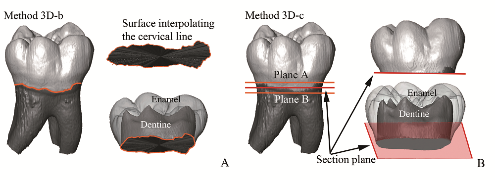
In computer-aided dental anthropology it is sometimes a regular process to separate the crown from the roots. In order to assess the methodological impact of sectioning crown and roots for the computation of enamel thickness, we compared two digital approaches(separating the crown from the root using the cervical line or a basal plane) for the 3D analysis of enamel thickness on a total number of 82 hominin lower postcanine teeth, including South African fossil hominins(n=26), Neanderthals(n=22), and modern humans(n=34). According to paired t-test, no significant difference is observed in the enamel thickness values between two methods, but subsequent inter-taxa comparisons reveal different results in average enamel thickness(AET) in premolars. Separation based on a basal plane is more operator-dependent, not practical to sinuous cervical margin and might mask between-group distinctions. Besides providing a set of raw data for further investigation, this study reports thinner premolar RET in Neanderthals compared with modern H. sapiens and therefore support the notion that Neanderthal has generally thinner relative enamel. Our results show that, for studies aimed at discriminating among different species, using the cervical margin to isolate the crown from the root is a practical option as it considers the anatomical nature of tooth, especially for those specimens(such as anterior dentition, or molars of Pan and Gorilla) with steep cervical line.

Lei PAN . Effects of two separation methods of crown and root on enamel thickness measurements[J]. Acta Anthropologica Sinica, 2019 , 38(03) : 398 -406 . DOI: 10.16359/j.cnki.cn11-1963/q.2018.0028
| [1] | Kay R. The nut-crackers—A new theory of the adaptations of the Ramapithecinae[J]. American Journal of Physical Anthropology, 1981,55:141-151 |
| [2] | Kay R. Dental evidence for the diet of Australopithecus[J]. Annual Review of Anthropology, 1985,14:315-341 |
| [3] | Martin L. Significance of enamel thickness in hominoid evolution[J]. Nature, 1985,314:260-263 |
| [4] | Ungar PS, Grine FE, Teaford MF, et al. Dental microwear and diets of African early Homo[J]. Journal of Human Evolution, 2006,50:78-95 |
| [5] | Kono R, Suwa G. Enamel distribution patterns of extant human and hominoid molars: occlusal versus lateral enamel thickness[J]. Bulletin of the National Museum of Nature and Science, 2008,34:1-9 |
| [6] | Olejniczak A, Tafforeau P, Feeney RNM, et al. Three-dimensional primate molar enamel thickness[J]. Journal of Human Evolution, 2008,54:187-195 |
| [7] | Smith TM, Olejniczak AJ, Reh S, et al. Brief communication: Enamel thickness trends in the dental arcade of humans and chimpanzees[J]. American Journal of Physical Anthropology, 2008,136:237-241 |
| [8] | Beynon A, Wood B. Variations in enamel thickness and structure in east African hominids[J]. American Journal of Physical Anthropology, 1986,70:177-193 |
| [9] | White TD, Suwa G, Asfaw B. Australopithecus ramidus, a new species of early hominid from Aramis, Ethiopia[J]. Nature, 1994,371:306-312 |
| [10] | Molnar S, Hildebolt C, Molnar IM, et al. Hominid enamel thickness: I. The Krapina neandertals[J]. American Journal of Physical Anthropology, 1993,92:131-138 |
| [11] | Smith TM, Olejniczak AJ, Zermeno JP, et al. Variation in enamel thickness within the genus Homo[J]. Journal of Human Evolution, 2012,62:395-411 |
| [12] | Skinner MM, Alemseged Z, Gaunitz C, et al. Enamel thickness trends in Plio-Pleistocene hominin mandibular molars[J]. Journal of Human Evolution, 2015,85:35-45 |
| [13] | Schwartz GT. Taxonomic and functional aspects of enamel cap structure in South African plio-pleistocene hominids: a high resolution computed tomographic study[D]. Ph. D. Dissertation, Washington University, 1997, 1-22 |
| [14] | Zhang LZ, Zhao LX. Enamel thickness of Gigantopithecus blacki and its significance for dietary adaptation and phylogeny[J]. Acta Anthropologica Sinica, 2013,32:365-376 |
| [15] | Tafforeau P. Phylogenetic and functional aspects of tooth enamel microstructure and three-dimensional structure of modern and fossil primate molars[D]. Ph.D. Dissertation, Université de Montpellier II, 2004, 1-133 |
| [16] | Olejniczak A. Micro-computed tomography of primate molars[D]. Ph. D. Dissertation, Stony Brook University, 2006, 1-194 |
| [17] | Feeney RNM, Zermeno JP, Reid DJ, et al. Enamel thickness in Asian human canines and premolars[J]. Anthropological Science, 2010,118:191-198 |
| [18] | Benazzi S, Panetta D, Fornai C, et al. Technical Note: Guidelines for the digital computation of 2D and 3D enamel thickness in hominoid teeth[J]. American Journal of Physical Anthropology, 2014,153:305-313 |
| [19] | Benazzi S, Fornai C, Bayle P, et al. Comparison of dental measurement systems for taxonomic assignment of Neanderthal and modern human lower second deciduous molars[J]. Journal of Human Evolution, 2011,61:320-326 |
| [20] | Fiorenza L, Benazzi S, Tausch J, et al. Molar macrowear reveals Neanderthal eco-geographic dietary variation[J]. PLOS ONE, 2011,6:e14769 |
| [21] | Beaudet A, Dumoncel J, Thackeray F, et al. Upper third molar internal structural organization and semicircular canal morphology in Plio-Pleistocene South African cercopithecoids[J]. Journal of Human Evolution, 2016,95:104-120 |
| [22] | Olejniczak A, Smith TM, Feeney RNM, et al. Dental tissue proportions and enamel thickness in Neandertal and modern human molars[J]. Journal of Human Evolution, 2008,55:12-23 |
| [23] | Zanolli C. Brief communication: molar crown inner structural organization in Javanese Homo erectus[J]. American Journal of Physical Anthropology, 2015,156:148-157 |
| [24] | Kuman K, Clarke R. Stratigraphy, artefacts, industries and hominid associations for Sterkfontein, Member 5[J]. Journal of Human Evolution, 2000,38:827-847 |
| [25] | Balter V, Blichert-Toftv J, Braga J, et al. U-Pb dating of fossil enamel from the Swartkrans Pleistocene hominid site, South Africa[J]. Earth and Planetary Science Letters, 2008,267:236-246 |
| [26] | Rink WJ, Schwarcz HP, Smith FH, et al. ESR dates for Krapina hominids[J]. Nature, 1995,378:24 |
| [27] | Girard M. La brèche à “Machairodus” de Montmaurin(Pyrénées centrales)[J]. Bulletin de l’Association Fran?aise pour l’étude du Quaternaire, 1973,3:193-207 |
| [28] | Macchiarelli R, Bondioli L, Debénath A, et al. How Neanderthal molar teeth grew[J]. Nature, 2006,444:748-751 |
| [29] | Pan L, Dumoncel J, de Beer F, et al. Further morphological evidence on South African earliest Homo lower postcanine dentition: enamel thickness and enamel dentine junction[J]. Journal of Human Evolution, 2016,96:82-96 |
| [30] | Molnar S. Human tooth wear, tooth function and cultural variability[J]. American Journal of Physical Anthropology, 1971,34:175-189 |
| [31] | Kono R. Molar enamel thickness and distribution patterns in extant great apes and humans, new insights based on a 3-dimensional whole crown perspective[J]. Anthropological Science, 2004,112:121-146 |
| [32] | Buti L, Le Cabec A, Panetta D, et al. 3D enamel thickness in Neandertal and modern human permanent canines[J]. Journal of Human Evolution, 2017,113:162-172 |
| [33] | Benazzi S, Slon V, Talamo S, et al. The makers of the Protoaurignacian and implications for Neandertal extinction[J]. Science, 2015,348:793-796 |
| [34] | Kono R, Suwa G, Tanijiri T. A three-dimensional analysis of enamel distribution patterns in human permanent first molars[J]. Archives of Oral Biology, 2002,47:867-875 |
| [35] | Kono RT, Zhang Y, Jin C, et al. A 3-dimensional assessment of molar enamel thickness and distribution pattern in Gigantopithecus blacki[J]. Quaternary International, 2014,354:46-51 |
| [36] | Zanolli C, Pan L, Dumoncel J, et al. Inner tooth morphology of Homo erectus from Zhoukoudian. New evidence from an old collection housed at Uppsala University, Sweden[J]. Journal of Human Evolution, 2018,116:1-13 |
/
| 〈 |
|
〉 |