
Received date: 2019-05-28
Revised date: 2019-06-18
Online published: 2020-09-10
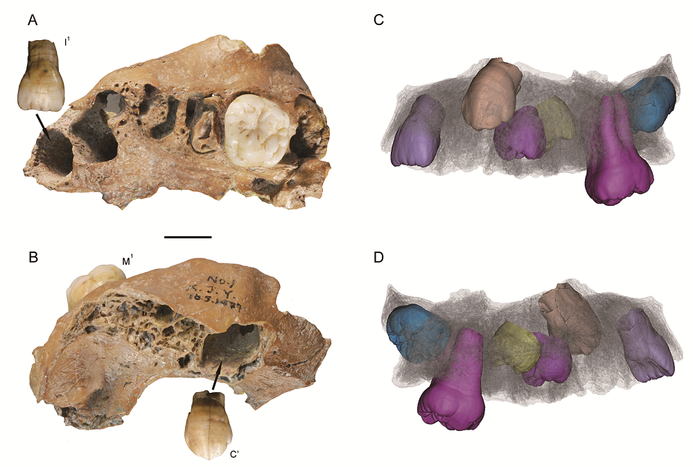
The hominin fossils recovered from Xujiayao-Hsuchiayao (Locality 74093)site is critical in understanding the morphological variability of hominins from the period of Middle to late Pleistocene transition. Other than the morphologies, the pathological aspects of Xujiayao hominins were also investigated and the juvenile (Xujiayao 1) was believed to be suffered from dental fluorosis based on the presence of the yellow pit or furrow on its anterior teeth. The incidence of “dental fluorosis” in Xujiayao has been thought to represent the earliest evidence of this pathological anomaly. However, with the use of scanning electron microscopy (SEM) and microcomputed tomography (micro-CT), the yellow enamel defects were found to be hypoplastic alternation that occurred before the tooth eruption. They were not post-eruptive physical breakage resulted from chewing force and therefore don’t support the diagnosis of dental fluorosis. In addition, synchrotron phase-contrast microtomography of the anterior teeth under both micron and submicron resolutions didn’t show obvious sign of subsurface hypomineralization along the sagittal section of the enamel, and it doesn’t support the occurrence of dental fluorosis resulted from the disturbance of enamel maturation phase. However, plenty of pit-type hypoplastic defects were present on the enamel surface of the Xujiayao permanent teeth and the bottom of the enamel pits were underlain by accentuated incremental line. This type of enamel defects could be resulted from distributed secretory stage of enamel formation by excessive fluoride intake according to the experimental study on mammals. Apart from the surface defects, synchrotron scanning under submicron resolution at four different spots of Xujiayao permanent teeth reveals plenty of enamel holes inside the crown. These holes have a sphere-like shape and generally restricted to outer one-third area of the enamel thickness. Different locations within one scanning spot vary in the density of enamel holes. In canine, the enamel holes of high-density correspond with a hypoplastic depression on the enamel surface. Enamel holes inside the paracone apex of m1 are in some cases connected with each other, with the main axis perpendicular to the outer enamel surface. These characteristics indicate a non-random distribution pattern of the enamel holes, and that they might be caused by the same effects as that of enamel hypoplasia. Teeth forming at different times vary in the density of hypoplastic pits and enamel holes, and this might imply various level of physiological disturbance at different stages of dental growth and development. Future study could further quantify the fluoride content in the deposit containing the Xujiayao hominin fossils, as well as in the tooth enamel, in order to ascertain if the Xujiayao people used to live a fluoride-rich environment and if they did ingest enough fluoride. With this information, the mechanism of enamel defects in Xujiayao juvenile could be more thoroughly understood.

Key words: Xujiayao hominin; Dental fluorosis; Synchrotron; Enamel defect
Song XING . Enamel defects of the Xujiayao juvenile[J]. Acta Anthropologica Sinica, 2019 , 38(04) : 499 -512 . DOI: 10.16359/j.cnki.cn11-1963/q.2019.0049
| [1] | 贾兰坡, 卫奇, 李超荣. 许家窑旧石器时代文化遗址-1976 年发掘报告[J]. 古脊椎动物与古人类, 1979,17:277-293 |
| [2] | 吴茂霖. 许家窑遗址1977 年出土的人类化石[J]. 古脊椎动物与古人类, 1980,18:229-238 |
| [3] | 吴茂霖. 许家窑人颞骨研究[J]. 人类学学报, 1986,5:220-226 |
| [4] | 陈铁梅, 原思训, 高世君. 铀子系法测定骨化石年龄的可靠性研究及华北地区主要旧石器地点的铀子系年代序列[J]. 人类学学报, 1984,3:259-269 |
| [5] | Nagatomo T, Shitaoka Y, Namioka H, et al. OSL dating of the strata at Paleolithic sites in the Nihewan Basin, China[J]. Acta Anthropologica Sinica, 2009,28:276-284 |
| [6] | Li Z, Xu Q, Zhang S, Hun L, et al. Study on stratigraphic age, climate changes and environment background of Houjiayao Site in Nihewan Basin[J]. Quaternary international, 2014,349:42-48 |
| [7] | Tu H, Shen G, Li H, et al. 26Al/10Be burial dating of Xujiayao-Houjiayao site in Nihewan Basin, northern China[J]. PLoS ONE, 2015,10, e0118315 |
| [8] | Ao H, Liu CR, Roberts AP, et al. An updated age for the Xujiayao hominin from the Nihewan Basin, North China: Implications for Middle Pleistocene human evolution in East Asia[J]. Journal of Human Evolution, 2017,106:54-65 |
| [9] | 王法岗, 李锋. “许家窑人”埋藏地层与时代探讨[J]. 人类学学报, 2019,38(e). DOI: 10.16359/j.cnki.cn11-1963/q.2019.0025 |
| [10] | Wu XJ, Crevecoeur I, Liu W, et al. Temporal labyrinths of eastern Eurasian Pleistocene humans[J]. Proceedings of the National Academy of Sciences of the United States of America, 2014,111:10509-10513 |
| [11] | Wu XJ, Trinkaus E. The Xujiayao 14 mandibular ramus and Pleistocene Homo mandibular variation[J]. Comptes Rendus Palevol, 2014,13:333-341 |
| [12] | Xing S, Martinón-Torres M, Bermúdez de Castro JM, et al. Hominin teeth from the early Late Pleistocene site of Xujiayao, Northern China[J]. American Journal of Physical Anthropology, 2015,156:224-240 |
| [13] | Wu XJ, Xing S, Trinkaus E. An enlarged parietal foramen in the late archaic Xujiayao 11 neurocranium from northern China, and rare anomalies among Pleistocene Homo[J]. PLoS ONE, 2013,8:59587 |
| [14] | Wu XJ, Trinkaus E. Neurocranial trauma in the late archaic human remains from Xujiayao, Northern China[J]. International Journal of Osteoarchaeology, 2015,25:245-252 |
| [15] | Xing S, Guatelli-Steinberg D, O’Hara M, et al. Micro-CT Imaging and Analysis of Enamel Defects on the Early Late Pleistocene Xujiayao Juvenile[J]. International Journal of Osteoarchaeology, 2016,26:935-946 |
| [16] | 李森照, 卫奇. 从 “许家窑人” 的牙病说起[J]. 化学通报, 1980,12:9 |
| [17] | Hillson S, Bond S. Relationship of enamel hypoplasia to the pattern of tooth crown growth: a discussion[J]. American Journal of Physical Anthropology: The Official Publication of the American Association of Physical Anthropologists, 1997,104:89-103 |
| [18] | Fejerskov O, Johnson NW, Silverstone LM. The ultrastructure of fluorosed human dental enamel[J]. European Journal of Oral Sciences, 1974,82:357-372 |
| [19] | Fejerskov O, Thylstrup A, Larsen MJ. Clinical and structural features and possible pathogenic mechanisms of dental fluorosis[J]. European Journal of Oral Sciences, 1977,85:510-534 |
| [20] | Fejerskov O, Yanagisawa T, Tohda H, et al. Posteruptive changes in human dental fluorosis--a histological and ultrastructural study[J]. Proceedings of the Finnish Dental Society. Suomen Hammaslaakariseuran toimituksia, 1991,87:607-619 |
| [21] | DenBesten PK Crenshaw MA. The effects of chronic high fluoride levels on forming enamel in the rat[J]. Archives of oral biology, 1984,29:675-679 |
| [22] | Aoba T, Fejerskov O. Dental fluorosis: chemistry and biology[J]. Critical Reviews in Oral Biology & Medicine, 2002,13:155-170 |
| [23] | DenBesten P, Li W. Chronic fluoride toxicity: dental fluorosis[J]. Fluoride and the oral environment, 2011,22:81-96 |
| [24] | Thylstrup A, Fejerskov O. Clinical appearance of dental fluorosis in permanent teeth in relation to histologic changes[J]. Community dentistry and oral epidemiology, 1978,6:315-328 |
| [25] | Dean HT. Fluorine in the control of dental caries. The Journal of the American Dental Association, 1956,52:1-8 |
| [26] | Hillson SW. Dental Anthropology[M]. Cambridge: Cambridge University Press, 1996 |
| [27] | Walton RE, Eisenmann DR. Ultrastructural examination of various stages of amelogenesis in the rat following parenteral fluoride administration[J]. Archives of oral biology, 1974, 19:171-IN17 |
| [28] | Monsour PA, Harbrow DJ, Warshawsky H. Effects of acute doses of sodium fluoride on the morphology and the detectable calcium associated with secretory ameloblasts in rat incisors[J]. Journal of Histochemistry & Cytochemistry, 1989,37:463-471 |
| [29] | Kierdorf H, Kierdorf U. Disturbances of the secretory stage of amelogenesis in fluorosed deer teeth: a scanning electron-microscopic study[J]. Cell and tissue research, 1997,289:125-135 |
| [30] | Kierdorf H, Kierdorf U, Richards A, et al. Fluoride-induced alterations of enamel structure: an experimental study in the miniature pig[J]. Anatomy and embryology, 2004,207:463-474 |
| [31] | Witzel C, Kierdorf U, Schultz M, et al. Insights from the inside: Histological analysis of abnormal enamel microstructure associated with hypoplastic enamel defects in human teeth[J]. American Journal of Physical Anthropology, 2008,136:400-414 |
| [32] | Kierdorf H, Kierdorf U, Richards A, et al. Disturbed enamel formation in wild boars(Sus scrofa L.) from fluoride polluted areas in Central Europe[J]. The Anatomical Record: An Official Publication of the American Association of Anatomists, 2000,259:12-24 |
| [33] | DenBesten PK. Dental fluorosis: its use as a biomarker[J]. Advances in dental research, 1994,8:105-110 |
| [34] | Whitford GM. Intake and metabolism of fluoride[J]. Advances in dental research, 1994,8:5-14 |
/
| 〈 |
|
〉 |