
Enamel-dentine junction shape and enamel thickness distribution of East Asian Middle Pleistocene hominin lower second molars
Received date: 2020-03-27
Revised date: 2020-04-30
Online published: 2020-09-11
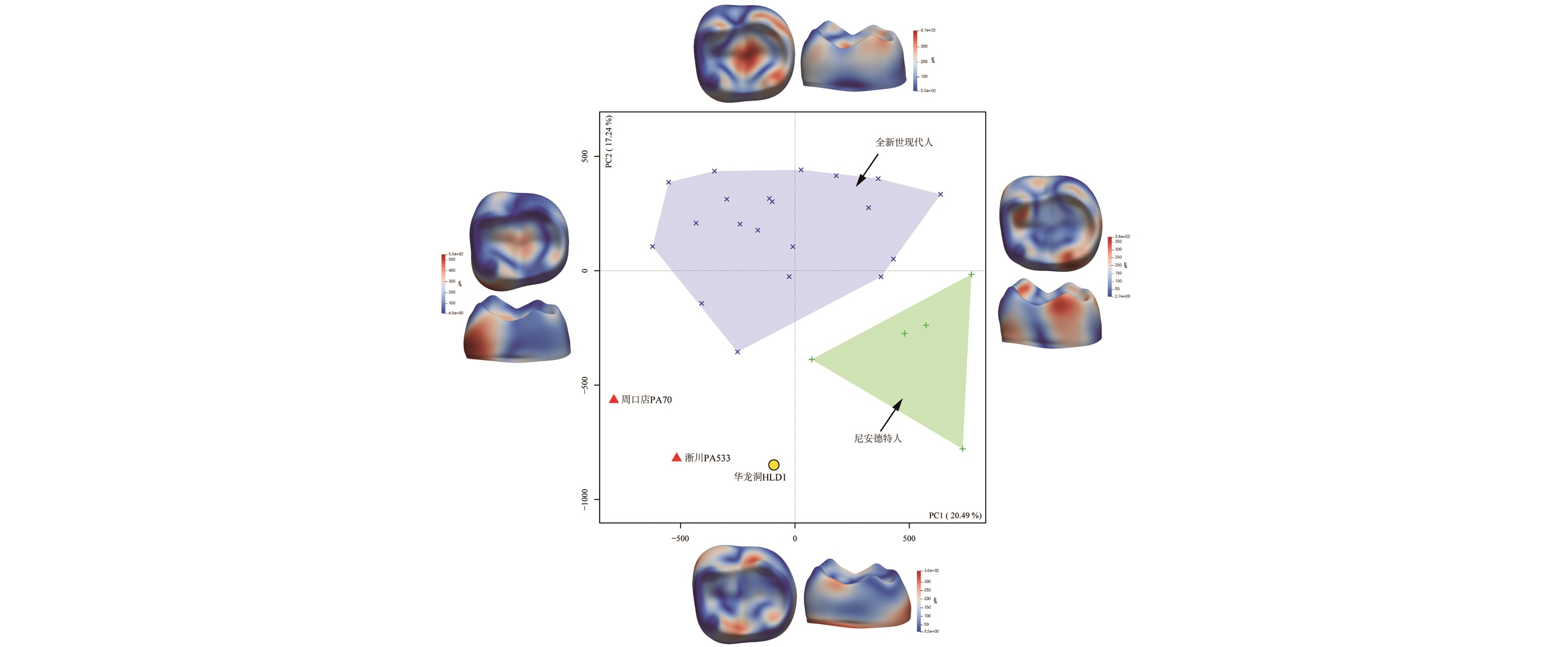
Morphological diversity has been revealed in the cranial, mandibular, and dental materials of East Asian Middle Pleistocene hominins, and the taxonomy of later members is uncertain. In order to further apprehend the morphological variability of East Asian Middle Pleistocene hominins and provide evidence for the taxonomy of the later members, the present study conducted three-dimensional morphometric analyses, including morphometric map of lateral enamel thickness and diffeomorphic surface matching of enamel-dentine junction (EDJ), on the lower second molars. The results indicate that: 1) East Asian Middle Pleistocene hominins could be distinguished from both Neanderthals and modern human; 2) East Asian late Middle Pleistocene hominins displayed special distribution pattern of lateral enamel thickness and a more progressive EDJ shape relative to mid-Middle Pleistocene Homo erectus. The present study quantifies two important morphologies and their variability, i.e., distribution pattern of enamel thickness and EDJ shape, in addition to the individual dental traits studied by previous works. It will provide further insight into the taxonomies of East Asian late Middle Pleistocene hominins and help designating isolated teeth from the same period into their correct morphological groups.

Key words: East Asian hominins; Teeth; Dentine; Three-dimensional morphometric
Song XING , Mi ZHOU , Lei PAN . Enamel-dentine junction shape and enamel thickness distribution of East Asian Middle Pleistocene hominin lower second molars[J]. Acta Anthropologica Sinica, 2020 , 39(04) : 521 -531 . DOI: 10.16359/j.cnki.cn11-1963/q.2020.0019
| [1] | Wu X, Poirier FE. Human evolution in China: a metric description of the fossils and a review of the sites[M]. Oxford University Press, USA, 1995 |
| [2] | 刘武, 吴秀杰, 邢松, 等. 中国古人类化石[M]. 北京: 科学出版社, 2014 |
| [3] | 刘武, 吴秀杰, 邢松. 更新世中期中国古人类演化区域连续性与多样性的化石证据[J]. 人类学学报, 2019,38:473-490 |
| [4] | Li ZY, Wu XJ, Zhou LP, et al. Late Pleistocene archaic human crania from Xuchang, China[J]. Science, 2017,355:969-972 |
| [5] | Buck LT, Stringer CB. Homo heidelbergensis[J]. Current Biology, 2014,24:R214-R215 |
| [6] | Stringer CB, Barnes I. Deciphering the Denisovans[J]. Proceedings of the National Academy of Sciences, USA, 2015,112:15542-15543 |
| [7] | Wu XJ, Maddux SD, Pan LEI, et al. Nasal floor variation among eastern Eurasian Pleistocene Homo[J]. Anthropological Science, 2012,120:217-226 |
| [8] | Gokhman D, Mishol N, de Manuel M, et al. Reconstructing Denisovan anatomy using DNA methylation maps[J]. Cell, 2019, 179: 180-192.e110 |
| [9] | Liu W, Zhang Y, Wu X. Middle Pleistocene human cranium from Tangshan (Nanjing), Southeast China: A new reconstruction and comparisons with Homo erectus from Eurasia and Africa[J]. American Journal of Physical Anthropology, 2005,127:253-262 |
| [10] | Cui Y, Wu X. A geometric morphometric study of a Middle Pleistocene cranium from Hexian, China[J]. Journal of Human Evolution, 2015,88:54-69 |
| [11] | Liu W, Martinón-Torres M, Kaifu Y, et al. A mandible from the Middle Pleistocene Hexian site and its significance in relation to the variability of Asian Homo erectus[J]. American Journal of Physical Anthropology, 2017,162:715-731 |
| [12] | Liu W, Schepartz LA, Xing S, et al. Late Middle Pleistocene hominin teeth from Panxian Dadong, South China[J]. Journal of Human Evolution, 2013,64:337-355 |
| [13] | Xing S, Martinón-Torres M, Bermúdez de Castro JM, et al. Hominin teeth from the early Late Pleistocene site of Xujiayao, Northern China[J]. American Journal of Physical Anthropology, 2015,156:224-240 |
| [14] | Xing S, Martinón-Torres M, de Castro JMB, et al. Middle Pleistocene hominin teeth from Longtan Cave, Hexian, China[J]. PLOS ONE, 2014,9:e114265 |
| [15] | Xing S, Sun C, Martinón-Torres M, et al. Hominin teeth from the Middle Pleistocene site of Yiyuan, Eastern China[J]. Journal of Human Evolution, 2016,95:33-54 |
| [16] | Xing S, Martinón-Torres M, Bermúdez de Castro JM. The fossil teeth of the Peking Man[J]. Scientific Reports, 2018,8:1-11 |
| [17] | Xing S, Martinón-Torres M, Bermúdez de Castro JM. Late Middle Pleistocene hominin teeth from Tongzi, southern China[J]. Journal of Human Evolution, 2019,130:96-108 |
| [18] | Xing S, Martinón-Torres M, Bermúdez de Castro JM. et al. Middle Pleistocene Hominin Teeth from Longtan Cave, Hexian, China[J]. PLOS ONE, 2015,9:e114265 |
| [19] | Butler P. The ontogeny of molar pattern[J]. Biological Reviews, 1956,31:30-69 |
| [20] | Skinner MM, Gunz P, Wood BA, et al. Enamel-dentine junction (EDJ) morphology distinguishes the lower molars of Australopithecus africanus and Paranthropus robustus[J]. Journal of Human Evolution, 2008,55:979-988 |
| [21] | Skinner MM, Wood B, Hublin JJ. Protostylid expression at the enamel-dentine junction and enamel surface of mandibular molars of Paranthropus robustus and Australopithecus africanus[J]. Journal of Human Evolution, 2009,56:76-85 |
| [22] | 刘武, 周蜜, 邢松. 卡氏尖在中国古人类化石中出现及其演化意义[J]. 人类学学报, 2018,37:159-175 |
| [23] | Pan L, Dumoncel J, de Beer F, et al. Further morphological evidence on South African earliest Homo lower postcanine dentition: enamel thickness and enamel dentine junction[J]. Journal of Human Evolution, 2016,96:82-96 |
| [24] | Zanolli C, Kullmer O, Kelley J, et al. Evidence for increased hominid diversity in the Early-Middle Pleistocene of Java, Indonesia[J]. Nature Ecology & Evolution, 2019,3:755-764 |
| [25] | Zanolli C, Martinón-Torres M, Bernardini F, et al. The Middle Pleistocene (MIS 12) human dental remains from Fontana Ranuccio (Latium) and Visogliano (Friuli-Venezia Giulia), Italy. A comparative high resolution endostructural assessment[J]. PLOS ONE, 2018,13:e0189773 |
| [26] | Olejniczak A, Smith TM, Skinner MM, et al. Three-dimensional molar enamel distribution and thickness in Australopithecus and Paranthropus[J]. Biology Letters, 2008,4:406-410 |
| [27] | Smith TM, Olejniczak AJ, Zermeno JP, et al. Variation in enamel thickness within the genus Homo[J]. Journal of Human Evolution, 2012,62:395-411 |
| [28] | Braga J, Zimmer V, Dumoncel J, et al. Efficacy of diffeomorphic surface matching and 3D geometric morphometrics for taxonomic discrimination of Early Pleistocene hominin mandibular molars[J]. Journal of Human Evolution, 2019,130:21-35 |
| [29] | Durrleman S, Pennec X, Trouvé A, et al. Comparison of the endocranial ontogenies between chimpanzees and bonobos via temporal regression and spatiotemporal registration[J]. Journal of Human Evolution, 2012,62:74-88 |
| [30] | Beaudet A, Dumoncel J, de Beer F, et al. Morphoarchitectural variation in South African fossil cercopithecoid endocasts[J]. Journal of Human Evolution, 2016,101:65-78 |
| [31] | Beaudet A, Dumoncel J, Thackeray F, et al. Upper third molar internal structural organization and semicircular canal morphology in Plio-Pleistocene South African cercopithecoids[J]. Journal of Human Evolution, 2016,95:104-120 |
| [32] | Urciuoli A, Zanolli C, Beaudet A, et al. The evolution of the vestibular apparatus in apes and humans[J]. eLife, 2020,9:e51261 |
| [33] | Zanolli C, Pan L, Dumoncel J, et al. Inner tooth morphology of Homo erectus from Zhoukoudian. New evidence from an old collection housed at Uppsala University, Sweden[J]. Journal of Human Evolution, 2018,116:1-13 |
| [34] | Morimoto N, De León MSP, Zollikofer CPE. Exploring femoral diaphyseal shape variation in wild and captive chimpanzees by means of morphometric mapping: A test of Wolff's law[J]. The Anatomical Record, 2011,294:589-609 |
| [35] | Puymerail L, Ruff CB, Bondioli L, et al. Structural analysis of the Kresna 11 Homo erectus femoral shaft (Sangiran, Java)[J]. Journal of Human Evolution, 2012,63:741-749 |
| [36] | Puymerail L, Volpato V, Debénath A, et al. A Neanderthal partial femoral diaphysis from the “grotte de la Tour”, La Chaise-de-Vouthon (Charente, France): Outer morphology and endostructural organization[J]. Comptes Rendus Palevol, 2012,11:581-593 |
| [37] | Wei P, Wallace IJ, Jashashvili T, et al. Structural analysis of the femoral diaphyses of an early modern human from Tianyuan Cave, China[J]. Quaternary International, 2017,434:48-56 |
| [38] | Jashashvili T, Dowdeswell MR, Lebrun R, et al. Cortical structure of hallucal metatarsals and locomotor adaptations in hominoids[J]. PLOS ONE, 2015,10:e0117905 |
| [39] | Zanolli C, Bondioli L, Coppa A, et al. The late Early Pleistocene human dental remains from Uadi Aalad and Mulhuli-Amo (Buia), Eritrean Danakil: macromorphology and microstructure[J]. Journal of Human Evolution, 2014,74:96-113 |
| [40] | Molnar S. Human tooth wear, tooth function and cultural variability[J]. American Journal of Physical Anthropology, 1971,34:175-189 |
| [41] | NESPOS database. Neanderthal Studies Professional Online Service[DB/OL], 2019, https://www.nespos.org/display/openspace/Home |
| [42] | Dumoncel J, Durrleman S, Braga J, et al. Landmark-free 3D method for comparison of fossil hominins and hominids based on endocranium and EDJ shapes[J]. American Journal of Physical Anthropology, 2014, 153: suppl.56, 110 |
| [43] | Durrleman S, Pennec X, Trouvé A, et al. Comparison of the endocranial ontogenies between chimpanzees and bonobos via temporal regression and spatiotemporal registration[J]. Journal of Human Evolution, 2012,62:74-88 |
| [44] | Durrleman S, Prastawa M, Charon N, et al. Morphometry of anatomical shape complexes with dense deformations and sparse parameters[J]. NeuroImage, 2014,101:35-49 |
| [45] | Durrleman S, Prastawa M, Korenberg JR, et al. Topology preserving atlas construction from shape data without correspondence using sparse parameters[A]. In: Ayache, N, Delingette, H, Golland, P, Mori, K (Eds.), Proceedings of Medical image Computing and Computer Assisted Intervention[C]. Springer, Nice, France, 2012: 223-230 |
| [46] | Durrleman S, Prastawa M, Charon N, et al. Morphometry of anatomical shape complexes with dense deformations and sparse parameters[J]. NeuroImage, 2014,101:35-49 |
| [47] | B?ne A, Louis M, Martin B, et al. Deformetrica 4: An open-source software for statistical shape analysiss[A]//International Workshop on Shape in Medical Imaging[C]. Springer, Cham, 2018: 3-13 |
| [48] | R Development Core Team. R: A language and environment for statistical computing[EB/OL],. R Foundation for Statistical Computing, Vienna, 2012 |
| [49] | Dray S, Dufour AB. The ade4 package: Implementing the duality diagram for ecologists[J]. Journal of statistical software, 2007,22:1-20 |
| [50] | Schlager S, Profico A, Di Vincenzo F, et al. Retrodeformation of fossil specimens based on 3D bilateral semi-landmarks: Implementation in the R package “Morpho”[J]. PLOS ONE, 2018,13:e0194073 |
| [51] | Dumoncel J. add_colormap_shooting.py[CP/OL],, 2020, https://gitlab.com/jeandumoncel/tools-for-deformetrica/-/blob/master/src/postprocessing/add_colormap_shooting.py |
| [52] | Wu X-J, Pei S-W, Cai Y-J, et al. Archaic human remains from Hualongdong, China, and Middle Pleistocene human continuity and variation[J]. Proceedings of the National Academy of Sciences, USA, 2019,116:9820-9824 |
| [53] | Chen F, Welker F, Shen C-C, et al. A late Middle Pleistocene Denisovan mandible from the Tibetan Plateau[J]. Nature, 2019,569:409-412 |
| [54] | Pan L, Dumoncel J, Mazurier A, et al. Structural analysis of premolar roots in Middle Pleistocene hominins from China[J]. Journal of Human Evolution, 2019,136:102669 |
| [55] | Wu XJ, Trinkaus E. The Xujiayao 14 Mandibular Ramus and Pleistocene Homo Mandibular Variation[J]. Comptes Rendus Palevol, 2014,13:333-341 |
| [56] | Wu XJ, Crevecoeur I, Liu W, et al. Temporal labyrinths of eastern Eurasian Pleistocene humans[J]. Proceedings of the National Academy of Sciences, USA, 2014,111:10509-10513 |
/
| 〈 |
|
〉 |