
Classification and variation of the temporal line of skulls in modern human
Received date: 2021-04-19
Revised date: 2021-06-21
Online published: 2022-10-13
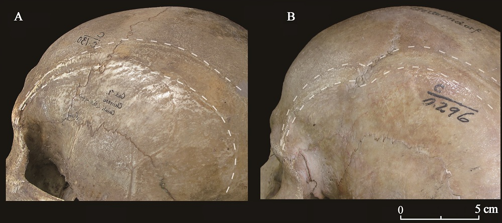
The temporal lines are muscle attachment marks bilaterally on the surface of the skull. Their morphological variations are meaningful to study the physical features and masticatory function in human evolution. In order to know the temporal line classifications and variations in modern populations, 278 adult skulls from Asia, Africa and Europe were selected to observe and analyze. Based on the arc trend, line width, development degree, roughness and terminal position, the temporal lines are classified into various types, and their identifications are standardly defined and established. The database of various temporal line types in sides, sexes and regions were obtained, that can be used for additional studies in physical anthropology. The primary results of this study show that: 1) there is no significant differences between left and right skull; 2) there were no significant differences in arc trend types and development degree between male and female, or among different regions; 3) There are sex difference in the width and roughness types, showing males have higher percentages than females; 4) There are spatial significant differences in the width and roughness types: Super-wide striped type was only found in the specimens from Yunnan and northern China. The Europeans have a high proportion of ridged type and rough types. The Africans have a high proportion of ridged type and a low proportion of rough type; 5) The specimens of temporal line ending in the occipital bone are very few; 6) The width on the frontal and parietal bones were significantly correlated with each other, as well as with the temporal line arc type; the development degree types on the frontal and parietal bones were significantly correlated with each other, too.

Yi YAN , Yuhao ZHAO , Xiujie WU . Classification and variation of the temporal line of skulls in modern human[J]. Acta Anthropologica Sinica, 2022 , 41(05) : 775 -787 . DOI: 10.16359/j.1000-3193/AAS.2021.0075
| [1] | Drake R, Vogl AW, Mitchell AWM. Gray’s anatomy for students (4th edition)[M]. London: Churchill Livingstone, 2019 |
| [2] | Robinson JT. Cranial cresting patterns and their significance in the Hominoidea[J]. American Journal of Physical Anthropology, 1958, 16(4): 397-428 |
| [3] | Laurin M, Reisz RR. Taxonomic position and phylogenetic relationships of Colobomycter pholeter, a small reptile from the Lower Permian of Oklahoma[J]. Canadian Journal of Earth Sciences, GeoScienceWorld, 1989, 26(3): 544-550 |
| [4] | Frazzetta TH. Adaptive problems and possibilities in the temporal fenestration of tetrapod skulls[J]. Journal of Morphology, 1968, 125(2): 145-157 |
| [5] | Gingerich PD, Boyer DM. Skeleton of Late Paleocene Plesiadapis Cookei (Mammalia, Euarchonta):Life History, Locomotion, and Phylogenetic Relationships[M]. Michigan:Museum of Paleontology, The University of Michigan, 2019 |
| [6] | Ashton EH, Zuckerman S. Cranial Crests in the Anthropoidea[J]. Proceedings of the Zoological Society of London, 1956, 126(4): 581-634 |
| [7] | Ahern JCM. Sahelanthropus[A]. In: Trevathan W (Ed.). The International Encyclopedia of Biological Anthropology[M]. American Cancer Society, Hoboken: Wiley-Blackwell, 2018: 1-6 |
| [8] | Potze S, Thackeray JF. Temporal lines and open sutures revealed on cranial bone adhering to matrix associated with Sts 5 (“Mrs Ples”), Sterkfontein, South Africa[J]. Journal of human evolution, 2010, 58(6): 533-535 |
| [9] | Laird MF, Schroeder L, Garvin HM, et al. The skull of Homo naledi[J]. Journal of Human Evolution, 2017, 104: 100-123 |
| [10] | Benazzi S, Gruppioni G, Strait DS, et al. Technical Note: Virtual reconstruction of KNM-ER 1813 Homo habilis cranium[J]. American Journal of Physical Anthropology, 2014, 153(1): 154-160 |
| [11] | Gabunia L, Vekua A, Lordkipanidze D, et al. Earliest Pleistocene Hominid Cranial Remains from Dmanisi, Republic of Georgia: Taxonomy, Geological Setting, and Age[J]. Science, 2000, 288(5468): 1019-1025 |
| [12] | Kaifu Y, Aziz F, Indriati E, et al. Cranial morphology of Javanese Homo erectus: New evidence for continuous evolution, specialization, and terminal extinction[J]. Journal of Human Evolution, 2008, 55(4): 551-580 |
| [13] | De Castro JMB, Martinón-Torres M, Arsuaga JL, et al. Twentieth anniversary of Homo antecessor (1997-2017): a review[J]. Evolutionary Anthropology: Issues News and Reviews, 2017, 26(4): 157-171 |
| [14] | Godinho RM, O’Higgins P. Chapter 10 - Virtual Reconstruction of Cranial Remains:The H. Heidelbergensis, Kabwe 1 Fossil[A]. In: Errickson D, Thompson T (Eds.). Human Remains: Another Dimension[M]. New York: Academic Press, 2017: 135-147 |
| [15] | Amano H, Kikuchi T, Morita Y, et al. Virtual reconstruction of the Neanderthal Amud 1 cranium[J]. American Journal of Physical Anthropology, 2015, 158(2): 185-197 |
| [16] | Argue D, Groves CP, Lee MSY, et al. The affinities of Homo floresiensis based on phylogenetic analyses of cranial, dental, and postcranial characters[J]. Journal of Human Evolution, 2017, 107: 107-133 |
| [17] | 吴新智, 席焕久, 陈昭. 人体测量手册[M]. 北京: 科学出版社, 2010 |
| [18] | 邰迎吉, 张炜, 李峻, 等. 增强的颞线——甲状旁腺机能亢进的一个骨改变征象[J]. 临床放射学杂志, 2003, 12(22): 1070 |
| [19] | Noback M L. Harvati KC. Covariation in the Human Masticatory Apparatus[J]. The Anatomical Record, 2015, 298(1): 64-84 |
| [20] | 加藤克知. 日本人頭蓋における側頭線,特にその発達度の時代差について[J]. 人類學雜誌, 1986, 94(4): 373-380 |
| [21] | 小倉信. インド人小児および成人頭蓋における下側頭線の計測学的研究[J]. 日本医科大学雑誌, 1986, 53(1): 21-34 |
/
| 〈 |
|
〉 |