
Non-metric traits of the human femoral head-neck junction
Received date: 2023-11-27
Revised date: 2024-01-08
Online published: 2024-06-04
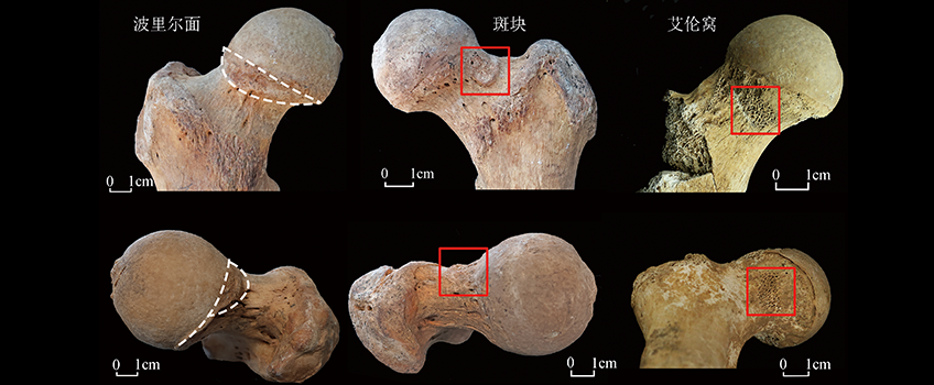
Various anatomical variations often occur on the anterior aspect of the femoral neck, and some features have been the subject of much research because of their possible relevance to ancient human behavior, such as Poirier’s facet. However, the definition of these non-metric traits and the reasons for their occurrence are controversial. In this paper, we combine the previous studies and practical observation experience to sort out the observation standards for the three common non-metric traits on the anterior femoral head-neck junction, namely, Poirier’s facet, plaque and Allen’s fossa. This paper observed eight groups of femur samples from archaeological sites in northern China dating from the Neolithic Age to the Ming-Qing period, and after statistics on the occurrence rate of each feature, we found that all three features showed significant gender, age, and population differences. Poirier’s facet and plaques were commonly found in males and occurred more frequently in middle-aged and older adults, and Allen’s fossa was more common in females and more prevalent in young adults. Combined with the anatomy of the hip joint and the daily activities of ancient populations, the presence of the Poirier’s facet and plaque may be associated with hip joint activity. Hip extension and external rotation increase the pressure on the iliofemoral ligament and compress the femoral head at the neck. With hip extension, external rotation and flexion, the femoral neck comes into contact with the acetabular rim, creating a pressure area. Femoroacetabular impingement is another possible factor that contributes to the appearance of Poirier’s facet and plaques. The formation of these two traits does not correlate well with behaviors such as riding horse and squatting. Allen’s fossa is different from both the morphological characteristics and the location of appearance of the Poirier’s facet and plaques, with Allen’s fossa appearing as early as childhood, possibly as a result of higher levels of stress in the individual’s survival, but more validation is needed from a sample of children. Taken together with the history of the research of Poirier facet and the reasons for its formation, the original translation of Poirier’s facet is not appropriate any longer. The significant differences in the performance of non-metric traits at the anterior aspects of the femoral head-neck junction between different populations suggest that there is potential for reconstructing activity patterns, health conditions and lifestyles of ancient people.

Key words: Poirier’s facet; riders’ facet; non-metric trait; femoral neck
CHENG Zhihan , CHONG Jianrong , SUN Zhanwei , YANG Lei , JING Xiaoting , WANG Jihong , HE Jianing . Non-metric traits of the human femoral head-neck junction[J]. Acta Anthropologica Sinica, 2024 , 43(03) : 415 -426 . DOI: 10.16359/j.1000-3193/AAS.2024.0034
| [1] | Singh I. Squatting facets on the talus and tibia in Indians[J]. Journal of Anatomy, 1959, 93(Pt 4): 540-550 |
| [2] | Ubelaker DH. Skeletal evidence for kneeling in prehistoric Ecuador[J]. American Journal of Physical Anthropology, 1979, 51(4): 679-685 |
| [3] | Angel JL. The reaction area of the femoral neck[J]. Clinical Orthopaedics and Related Research, 1964, 32: 130-142 |
| [4] | Charles RH. The Influence of Function, as Exemplified in the Morphology of the Lower Extremity of the Panjabi[J]. Journal of Anatomy and Physiology, 1893, 28(Pt 1): 1-18 |
| [5] | Pálfi Gy, Dutour O. Activity-induced skeletal markers in historical anthropological material[J]. International Journal of Anthropology, 1996, 11(1): 41-55 |
| [6] | Allen H. A system of human anatomy, including its medical and surgical relations[M]. Philadelphia: Henry C. Lea’s Son & Co, 1884 |
| [7] | 舒勒茨·米夏艾勒, 舒勒茨泰德·H·施米特, 巫新华, 等. 新疆于田县流水墓地26号墓出土人骨的古病理学和人类学初步研究[J]. 考古, 2008, 3: 86-91 |
| [8] | 魏东, 曾雯, 常喜恩, 等. 新疆哈密黑沟梁墓地出土人骨的创伤、病理及异常形态研究[J]. 人类学学报, 2012, 31(2): 176-186 |
| [9] | 原海兵, 秋吉尼玛索朗, 吕红亮, 等. 西藏那曲布塔雄曲青铜时代石室墓出土人骨研究[J]. 藏学学刊, 2017, 1: 273-300+321 |
| [10] | Molleson T, Blondiaux J. Riders’ Bones from Kish, Iraq[J]. Cambridge Archaeological Journal, 1994, 4(2): 312-316 |
| [11] | Khudaverdyan A, Khachatryan H, Eganyan L. Multiple trauma in a horse rider from the Late Iron Age cemetery at Shirakavan, Armenia[J]. Bioarchaeology of the near East, 2016, 12(10): 47-68 |
| [12] | Bühler B, Kirchengast S. A life on horseback? Prevalence and correlation of metric and non-metric traits of the “horse-riding syndrome” in an Avar population (7th-8th century AD) in Eastern Austria[J]. Anthropological Review, 2022, 85(3): 67-82 |
| [13] | 北京大学考古实习队, 河南省南阳市文物研究所. 河南邓州八里岗遗址发掘简报[J]. 文物, 1998, 9: 31-45+101 |
| [14] | 何嘉宁, 李楠, 张弛. 邓州八里岗仰韶时期居民的体质变迁[J]. 人类学学报, 2022, 41(4): 686-697 |
| [15] | 陕西省考古研究院. 2013年陕西省考古研究院考古发掘调查新收获[J]. 考古与文物, 2014, 2: 3-23+2+121 |
| [16] | 陕西省考古研究院. 2014年陕西省考古研究院考古调查发掘新收获[J]. 考古与文物, 2015, 2: 3-26+2+129 |
| [17] | 北京市文物研究所. 军都山墓地:玉皇庙(一)[M]. 北京: 文物出版社, 2007 |
| [18] | 何嘉宁, 唐小佳. 军都山古游牧人群股骨功能状况及流动性分析[J]. 科学通报, 2015, 60(17): 1612-1620 |
| [19] | 孙战伟, 夏培朝, 冯丹. 陕西洛川月家庄秦墓发掘简报[J]. 考古与文物, 2023, 1: 13-23 |
| [20] | 张海伦. 山西大同上华琚墓地人骨研究[D]. 硕士研究生毕业论文, 北京: 北京大学, 2023 |
| [21] | Radi N, Mariotti V, Riga A, et al. Variation of the anterior aspect of the femoral head-neck junction in a modern human identified skeletal collection[J]. American Journal of Physical Anthropology, 2013, 152(2): 261-272 |
| [22] | Henke W. Handbuch der Anatomie und Mechanik der Gelenke: mit Rücksicht auf Luxationen und Contracturen[M]. Leipzig: CF. Winter’sche Verlagshandlung, 1863 |
| [23] | Bertaux A. L’Humérus et le Fémur, considérés dans les Espèces dans les Races humaines selon le Sexe et selon l’Age[D]. Paris: A la Faculté de Médecine de Lille, 1891 |
| [24] | Poirier P, Charpy A. Traité d’anatomie humaine[M]. Paris: Masson, 1896 |
| [25] | Tramond E. Quelques particularités sur le fémur[D]. Paris: Faculté de Médecine de Prais, 1894 |
| [26] | Regnault F. Forme des surfaces articulaires des membres inférieurs[J]. Bulletins et Mémoires de la Société d’Anthropologie de Paris, 1898, 9(1): 535-544 |
| [27] | Pearson K, Bell J. A Study of the Long Bones of the English Skeleton[M]. Cambridge: Cambridge University Press, 1917 |
| [28] | Meyer AW. The genesis of the fossa of allen and associated structures[J]. American Journal of Anatomy, 1934, 55(3): 469-510 |
| [29] | Fick R. Handbuch der Anatomie und Mechanik der Gelenke[M]. Jena: Verlag Von Gustav Fisher, 1904 |
| [30] | Odgers PNB. Two Details about the Neck of the Femur. (1) The Eminentia. (2) The Empreinte[J]. Journal of Anatomy, 1931, 65(Pt 3): 352-362 |
| [31] | Finnegan M, Faust MA. Variants of the Femur[J]. Research of Report, 1974, 14: 7-20 |
| [32] | Finnegan M. Non-metric variation of the infracranial skeleton.[J]. Journal of Anatomy, 1978, 125(Pt 1): 23-37 |
| [33] | G?hring A. Allen’s fossa—An attempt to dissolve the confusion of different nonmetric variants on the anterior femoral neck[J]. International Journal of Osteoarchaeology, 2021, 31(4): 513-522 |
| [34] | 原海兵, 刘岩, 张光辉. 山西泽州县和村遗址出土春秋时期人骨初步研究[J]. 北方文物, 2017, 4: 51-53 |
| [35] | Kapandj AI. 骨关节功能解剖学:下肢(第6版)[M].译者:顾冬云, 戴尅戎. 北京: 人民军医出版社, 2015 |
| [36] | 姜卓蓝. 河南邓州八里岗古代人群蹲踞面初步研究[D]. 本科毕业论文, 北京: 北京大学, 2019 |
| [37] | Siebenrock KA, Wahab KHA, Werlen S, et al. Abnormal Extension of the Femoral Head Epiphysis as a Cause of Cam Impingement[J]. Clinical Orthopaedics and Related Research (1976-2007), 2004, 418: 54-60 |
| [38] | Ganz R, Parvizi J, Beck M, et al. Femoroacetabular Impingement: A Cause for Osteoarthritis of the Hip[J]. Clinical Orthopaedics and Related Research?, 2003, 417: 112 |
| [39] | J?ger M, Wild A, Westhoff B, et al. Femoroacetabular impingement caused by a femoral osseous head-neck bump deformity: clinical, radiological, and experimental results[J]. Journal of Orthopaedic Science, 2004, 9(3): 256-263 |
| [40] | Villotte S, Knüsel CJ. Some remarks about femoroacetabular impingement and osseous non-metric variations of the proximal femur[J]. Bulletins et mémoires de la Société d’Anthropologie de Paris, 2009, 21(1-2) |
| [41] | Steckel RH, Larsen CS, Sciulli PW, et al. Data collection codebook[A]. Steckel RH, Larsen CS, Roberts CA, et al(Eds). The Backbone of Europe: Health, Diet, Work, and Violence over Two Millennia[C]. Cambridge: Cambridge University Press, 2019 |
| [42] | Kostick EL. Facets and imprints on the upper and lower extremities of femora from a Western Nigerian population[J]. Journal of Anatomy, 1963, 97(Pt 3): 393-402 |
| [43] | Cunningham C, Scheuer L, Black S. Developmental Juvenile Osteology[M]. London: Academic Press, 2016 |
| [44] | Djuric M, Milovanovic P, Janovic A, et al. Porotic lesions in immature skeletons from Stara Torina, late medieval Serbia[J]. International Journal of Osteoarchaeology, 2008, 18(5): 458-475 |
| [45] | Trueta J, Harrison MHM. The normal vascular anatomy of the femoral head in adult man[J]. The Journal of Bone & Joint Surgery British Volume, 1953, 35-B(3): 442-461 |
| [46] | 高寿仙. 明代北京杂役考述[J]. 中国社会经济史研究, 2003, 4: 33-42 |
| [47] | 赵玉霞. 明长城蓟镇、昌镇、宣府镇和真保镇驿传系统研究[D]. 硕士研究生毕业论文, 天津: 天津大学, 2022 |
| [48] | 陈表义, 谭式玫. 明代军制建设原则及军事的衰败[J]. 暨南学报(哲学社会科学), 1996, 2: 58-65 |
| [49] | 袁靖. 中国动物考古学[M]. 北京: 文物出版社, 2015 |
| [50] | Berthon W, Tihanyi B, Kis L, et al. Horse riding and the shape of the acetabulum: Insights from the bioarchaeological analysis of early Hungarian mounted archers (10th century)[J]. International Journal of Osteoarchaeology, 2019, 29(1): 117-126 |
/
| 〈 |
|
〉 |