

Sex and age-at-death of the Yahuai Cave Man from the Late Pleistocene of Guangxi
Received date: 2024-01-05
Revised date: 2024-03-25
Online published: 2025-06-18
Obtaining accurate information about an individual’s sex and age at death constitutes the cornerstone of biological anthropological research. The estimation of adult sex from skeletal samples relies on two types of characteristics: those associated with functional differences between the sexes, and those manifested as variations in the size and shape of bones and teeth. Most methods for age estimation are based on the assessment of osteological degenerative changes. The identification of sex and age in human fossils often sparks controversy due to their evolutionary traits and preservation limitations, particularly when the hip bone is unavailable.
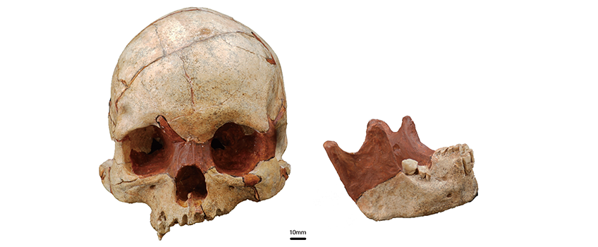
This study utilized visual observation, measurement, and micro-CT scanning to examine the Late Pleistocene crania, mandible, and teeth (YH1) excavated from the Yahuai Cave site in Guangxi, southern China. Regarding non-metric characteristics, the cranial and mandibular morphology indicates that Yahuai 1 is more likely to be female. However, certain robusticity-related features are more pronounced in YH1 compared to most recent-modern females in East Asia. The sexual dimorphism observed in late Pleistocene human skulls differs from that of modern humans, as evidenced by cranio-facial metric features. Comparisons with late Pleistocene human skulls from East and Southeast Asia reveal that YH1 shows no obvious sexual dimorphism in craniometric characteristics. When compared with recent populations from the same region, YH1 is found to be larger in size, and exhibits similarity to recent female samples after size calibration. YH1 has a medium-sized mandible overall and a high corpus robusticity index, which is comparable to that of late Pleistocene and Neolithic females.
In terms of age-at-death estimation, YH1 displayed a low degree of synostosis in the cranial sutures, and no age-associated features were observed. Moreover, by comparing with two Neolithic populations from South China, the relationship between dental wear and age estimation was adjusted. Collectively, these findings suggest that YH1 was not elderly, but rather a young adult aged between 30 and 40 years.
Key words: Yahuai Cave Man; sex; age-at-death; cranium; dental wear
HE Letian , XIE Guangmao , LIN Qiang , LI Dawei , WU Xiujie . Sex and age-at-death of the Yahuai Cave Man from the Late Pleistocene of Guangxi[J]. Acta Anthropologica Sinica, 2025 , 44(03) : 389 -403 . DOI: 10.16359/j.1000-3193/AAS.2025.0034
| [1] | Smith FH. Sexual differences in European Neanderthal crania with special reference to the Krapina Remains[J]. Journal of Human Evolution, 1980, 9(5): 359-375 |
| [2] | Franklin D, Cardini A, Flavel A, et al. Estimation of sex from cranial measurements in a Western Australian population[J]. Forensic Science International, 2013, 229(1-3): 158.e1-158.e8 |
| [3] | Clark MA, Cheverko CM, Simon A, et al. The decade under review: Recent trends and challenges in the use of macroscopic age-at-death estimation methods in bioarchaeology[J]. International Journal of Osteoarchaeology, 2023, 33(1): 150-163 |
| [4] | Phenice T. A newly developed visual method of sexing the OS pubis[J]. American Journal of Physical Anthropology, 1969, 30: 297-302 |
| [5] | Bruzek J. A method for visual determination of sex, using the human hip bone[J]. American Journal of Physical Anthropology, 2002, 117: 157-168 |
| [6] | Buikstra J, Ubelaker D(eds). Standards for data collection from human skeletal remains[M]. Arkansas Archaeological Survey: Fayetteville, 1994 |
| [7] | Iscan MY. Forensic anthropology of sex and body size[J]. Forensic Science International, 2005, 147: 107-112 |
| [8] | Ubelaker DH, DeGaglia CM. Population variation in skeletal sexual dimorphism[J]. Forensic Science International, 2017, 278: e401-e407 |
| [9] | Hassan NA-M, Brown KIA, Eyers J, et al. Ancient DNA study of the remains of putative infanticide victims from the Yewden Roman villa site at Hambleden, England[J]. Journal of Archaeological Science, 2014, 43: 192-197 |
| [10] | 张宝帅, 吴晓桐, 吴高, 等. 釉原蛋白微损分析方法鉴定人骨性别的有效性与优化[J]. 人类学学报, 2023, 42(4): 472-487 |
| [11] | Schmitt P, Murail E, Cunha DR. Variability of the pattern of aging on the human skeleton: evidence from bone indicators and implications on age at death estimation[J]. Journal of Forensic Sciences, 2002, 47(6): 1203-1209 |
| [12] | Xanthopoulou P, Valakos E, Youlatos D, et al. Assessing the accuracy of cranial and pelvic aging methods on human skeletal remains from a modern Greek assemblage[J]. Forensic Science International, 2018, 286: 266.e1-266.e8 |
| [13] | Brooks S, Suchey JM. Skeletal age determination based on the os pubis: A comparison of the Acsadi-Nemeskeri and Suchey-Brooks methods[J]. Journal of Human Evolution, 1990, 5, 227-238 |
| [14] | Meindl RS, Lovejoy CO. Ectocranial suture closure: A revised method for the determination of skeletal age at death based on the lateral-anterior sutures[J]. American Journal of Physical Anthropology, 1985, 68(1): 57-66 |
| [15] | Iscan MY, Loth SR, Wright RK. Age estimation from the rib by phase analysis- white males[J]. Journal of Forensic Sciences, 1984, 29(4): 1094-1104 |
| [16] | Iscan MY, Loth SR, Wright RK. Metamorphosis at the sternal rib end: A new method to estimate age at death in white males[J]. American Journal of Physical Anthropology, 1984, 65(2): 147-156 |
| [17] | Iscan MY, Loth SR, Wright RK. Age estimation from the rib by phase analysis white females[J]. Journal of Forensic Sciences, 1985, 30(3): 853-863 |
| [18] | Brothwell DR. The relationship of tooth wear to aging[A]. In: MY Is?an (ed.). Age Markers in the Human Skeleton[C]. Charles C Thomas Publisher, 1989, 303-316 |
| [19] | Lovejoy CO. Dental wear in the Libben population- its functional pattern and role in the determination of adult skeletal age at death[J]. American Journal of Physical Anthropology, 1985, 68(1): 47-56 |
| [20] | 魏博源, 朱文, 钟耳顺, 等. 广西崇左冲塘新石器遗址人骨和牙齿年龄的组织学鉴定[J]. 人类学学报, 1994, 2: 134-137 |
| [21] | Xing S, Tafforeau P, O’Hara M, et al. First systematic assessment of dental growth and development in an archaic hominin (genus, Homo) from East Asia[J]. Science Advance. 2019, 5(1): eaau0930 |
| [22] | Cerrito P, Nava A, Radov?i? D, et al. Dental cementum virtual histology of Neanderthal teeth from Krapina (Croatia, 130-120 kyr): an informed estimate of age, sex and adult stressors[J]. Journal of the Royal Society Interface, 2022, 19: 20210820 |
| [23] | Xu C, Qu H, Wang G, et al. A novel strategy for forensic age prediction by DNA methylation and support vector regression model[J]. Scientific Reports, 2015, 5: 17788 |
| [24] | Srettabunjong S, Satitsri S, Thongnoppakunw, et al. The study on telomere length for age estimation in a Thai population[J]. The American Journal of Forensic Medicine and Pathology, 2014, 35(2): 148-153 |
| [25] | Bolden JL, Milner GR, Konigsberg LW, et al. Transition analysis: a new method for estimating age from skeletons[A]. In: Hoppa RD,Vaupel JW(eds). Paleodemography: Age distributions from skeletal samples[C]. New York: Cambridge University Press, 2002, 73-106 |
| [26] | Getz SM. Improved skeletal age-at-death estimation and its impact on archaeological analyses(Doctoral Dissertation)[D]. University Park, PA: The Pennsylvania State University, 2017, 40-41 |
| [27] | Wu Y, Xie G, Mao L, et al. Phytolith evidence for human-plant subsistence in Yahuai Cave (Guangxi, South China) over the past 30000 years[J]. 中国科学:地球科学(英文版), 2020, 63(11): 1745-1757 |
| [28] | Matsumura H, Xie G, Nguyen LC, et al. Female craniometrics support the ‘two-layer model’ of human dispersal in Eastern Eurasia[J]. Scientific Report, 2021, 11: 20830 |
| [29] | 吴秀杰. 中国全新世人群头骨形态特征的变异[D].博士学位论文, 北京: 中国科学院研究生院, 2006 |
| [30] | Recommendations for age and sex diagnoses of skeleton[J]. Journal of Human Evolution, 1980, 9(7): 533-549 |
| [31] | 吴新智. 周口店山顶洞人化石的研究[J]. 古脊椎动物与古人类, 1961, 3: 181-211 |
| [32] | 吴汝康. 广西柳江发现的人类化石[J]. 古脊椎动物与古人类, 1959, 3: 97-104 |
| [33] | Storm P. The evolutionary significance of the Wajak skulls[M]. Nationaal Natuurhistorisch Museum, 1995 |
| [34] | Suzuki H. Skulls of Minatogawa Man[M]. University Museum Bulletin, The University of Tokyo, 1982, 19: 7-49 |
| [35] | Matsumura H, Yoneda M, Dodo Y, et al. Terminal Pleistocene human skeleton from Hang Cho cave, northern Vietnam: implications for the biological affinities of Hoabinhian people[J]. Anthropological Sciences. 2008, 116: 201-217 |
| [36] | Matsumura H, Pookajorn S. A morphometric analysis of the Late Pleistocene human skeleton from the Moh Khiew Cave in Thailand[J]. Homo, 2005, 56: 93-118 |
| [37] | Curnoe D, Datan I, Tacon PS, et al. Deep skull from Niah Cave and the Pleistocene peopling of Southeast Asia[J]. Frontiers in Ecology and Evolution, 2016, 4: 1-17 |
| [38] | Howells WW. Cranial variation in Man: A study by multivariate analysis of patterns of difference among recent human populations[M]. Cambridge: Harvard University Press, 1973 |
| [39] | Howells WW. Who’s Who in Skulls. Ethnic identification of crania from measurements[M]. Cambridge: Harvard University Press, 1995 |
| [40] | Howells WW. Howells’ Craniometric data on the Internet[J]. American Journal of Physical Anthropology. 1996, 101: 441-442 |
| [41] | Darroch JN, Mosimann JE. Canonical and principal components of shape[J]. Biometrika, 1985, 72: 241-252 |
| [42] | 李海军. 中国现代人群下颌骨的形态变异与功能适应[D]. 博士学位论文, 北京: 中国科学院研究生院, 2010 |
| [43] | Nikolova S, Toneva D, Georgiev I, et al. Sagittal suture maturation: morphological reorganization, relation to aging, and reliability as an age-at-death indicator[J]. American Journal of Physical Anthropology, 2019, 169(1): 78-92 |
| [44] | Fan F, Tu M, Li R, et al. Age estimation by multidetector computed tomography of cranial sutures in Chinese male adults[J]. American Journal of Physical Anthropology, 2020, 171(3): 550-558 |
| [45] | Chiba F, Makino Y, Motomura A, et al. Age estimation by multidetector CT images of the sagittal suture[J]. International Journal of Legal Medicine, 2013, 127: 1005-1011 |
| [46] | 莫世泰, 张文光, 雷绍伯, 等. 中国人颅骨缝的变化与年龄关系[J]. 人类学学报, 1989, 2: 131-138 |
| [47] | 裘诗文, 涂梦, 范飞, 等. 应用颅缝薄层CT扫描推断汉族成人年龄[J]. 法医学杂志, 2020, 36(4): 507-513 |
| [48] | Todd TW, Lyon D. Endocranial suture closure. Its progress and age relationship. Part I. adult males of white stock[J]. American Journal of Physical Anthropology, 1924, 7(3): 325-384 |
| [49] | Boldsen JL. Analysis of dental attrition and mortality in the Medieval village of Tirup, Denmark[J]. American Journal of Physical Anthropology, 2005, 126: 169-176 |
| [50] | Walker PL, Dean G, Shapiro P. Estimating age from tooth wear in archaeological populations[J]. Advances in Dental Anthropology, 1991, 169: 187 |
| [51] | McKee JK, Molnar S. Measurements of tooth wear among Australian aborigines: II. Intra-populational variation in patterns of dental attrition[J]. American Journal of Physical Anthropology, 1988, 76(1): 125-136 |
| [52] | 吴汝康, 柏惠英. 华北人颅骨臼齿磨耗年龄变化[J]. 古脊椎动物与古人类, 1965, 9(2): 217-222 |
| [53] | 莫世泰, 等. 对华南人颅骨臼齿磨耗年龄变化的研究[J]. 广西医学院学报, 1983, 1: 23-29 |
| [54] | Milella M, Franklin D, Belcastro MG, et al. Sexual differences in human cranial morphology: Is one sex more variable or one region more dimorphic?[J]. The Anatomical Record, 2021, 304: 2789-2810 |
| [55] | 杨茂有, 刘武, 邰凤久. 下颌骨的性别判别分析研究[J]. 人类学学报, 1988, 4: 329-334 |
| [56] | Harth S, Obert M, Ramsthaler F, et al. Estimating age by assessing the ossification degree of cranial sutures with the aid of Flat-Panel-CT[J]. Legal Medicine, 2009, 11(S1): s186-s189 |
| [57] | 国外刑事技术参考资料. 关于颅穹骨厚度的年龄变异性[J]. 刑事技术, 1977, 2: 33-36 |
| [58] | Boer D, Hans HH, Merwe VD, et al. Human cranial vault thickness in a contemporary sample of 1097 autopsy cases: relation to body weight, stature, age, sex and ancestry[J]. International Journal of Legal Medicine, 2016, 130(5): 1371-1377 |
| [59] | Elizabeth ML, Urban JE, Lynch SK, et al. Evaluation of Skull Cortical Thickness Changes With Age and Sex From Computed Tomography Scans[J]. Journal of Bone & Mineral Research, 2016, 32(2): 299-307 |
| [60] | 吴秀杰, 严毅. 资阳人头骨化石的内部解剖结构[J]. 人类学学报, 2020, 39(4): 511-520 |
| [61] | 吴汝康. 化石人类头骨年龄和性別的鉴定[J]. 古脊椎动物与古人类, 1962, 1: 116-118 |
| [62] | 张银运, 邢松. 周口店直立人3号头骨的性别和年龄:南京直立人头骨的启示[J]. 人类学学报, 2011, 30(3): 241-249 |
| [63] | Michel J, Paganelli A, Varoquaux A, et al. Determination of sex: interest of frontal sinus 3D reconstructions[J]. Journal of Forensic Sciences. 2015, 60(2): 269-273 |
| [64] | Zulkiflee NDI, Singh MKC, Alias A, et al. Morphological changes of the frontal sinus with age: a two-dimensional geometric morphometric study[J]. Forensic Imaging, 2023, 35: 200569 |
| [65] | Robles M, Rando C, Morgan RM. The utility of three-dimensional models of paranasal sinuses to establish age, sex, and ancestry across three modern populations: A preliminary study[J]. Australian Journal of Forensic Sciences, 2020, 54(3): 326-345 |
| [66] | Fatu C, Puisoru M, Rotaru M, et al. Morphometric evaluation of the frontal sinus in relation to age[J]. Annals of Anatomy-Anatomischer Anzeiger, 2006, 188(3): 275-280 |
| [67] | Genoves S. Sex determination in earlier man[A]. In: DBrothwell, EHiggs (eds). Science in Archaeology[M]. New York: Praeger, 1969, 342-352 |
| [68] | Keen JA. A study of sex differences in males and females skulls[J]. Americal Journal of Physical Anthropology, 1950, 8: 65-79 |
| [69] | Lahr MM, Wright RVS. The question of robusticity and the relationship between cranial size and shape in Homo sapiens[J]. Journal of Human Evolution, 1996, 31(2): 157-191 |
| [70] | 陈晓颖, 游海杰, 宋美玲, 等. 广西敢造遗址史前居民口腔的健康状况[J]. 人类学学报, 2023, 42(1): 98-109 |
| [71] | 陈伟驹, 李法军. 鲤鱼墩遗址出土人牙的牙齿磨耗和龋齿[J]. 人类学学报, 2013, 32(1): 45-51 |
| [72] | 中国社会科学院考古研究所, 等(编). 桂林甑皮岩[R]. 北京: 文物出版社, 2009, 405-425 |
| [73] | 朱芳武, 卢为善. 桂林甑皮岩新石器时代遗址2例儿童的年龄问题[J]. 人类学学报, 1995, 2: 147-150 |
| [74] | 张弛, 洪晓纯. 中国华南及其邻近地区的新石器时代采集渔猎文化[J]. 考古学研究, 2008, 415-434 |
| [75] | Armelagos GJ, Van Gerven DP. Sexual dimorphism and human evolution: an overview[J]. Journal of Human Evolution, 1980, 9: 437-446 |
/
| 〈 |
|
〉 |