
收稿日期: 2020-02-08
修回日期: 2020-04-10
网络出版日期: 2020-09-19
基金资助
中国科学院战略性先导科技专项(XDB26000000);国家自然科学基金(41802020);中国博士后科学基金面上项目(2017M611449)
Structural properties of the femoral remains from Lijiang, Yunnan province
Received date: 2020-02-08
Revised date: 2020-04-10
Online published: 2020-09-19
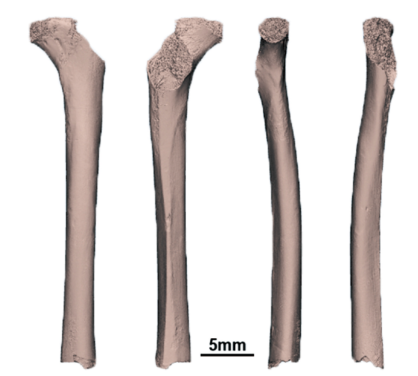
1960年,在云南省丽江市发现了三根古人类股骨,通过地层观察,仅PA108可归为更新世晚期。前人对PA108做了初步报导,为了进一步了解丽江人股骨的演化分类地位和东亚早期现代人股骨形态变异,本文对PA108的内外结构进行了详尽的分析。研究发现,PA108具有明显的早期现代人特征,即明显的股骨粗线、骨干中部后侧骨密质最厚和中部横断面轮廓形状偏椭圆。PA108标本也有一定的特殊性,体现在骨干中近端和中部骨密质厚度分布上,这可能与其股骨嵴发育较弱有关,这一特征也导致了PA108与其他东亚早期现代人之间的形态差异,这些形态变异进一步扩大了目前已知的东亚地区早期现代人变异范围。同时,在采用骨密质厚度分布模式进行分类时,建议关注股骨骨干中部骨密质最厚部位。

魏偏偏 . 云南丽江古人类股骨的形态结构[J]. 人类学学报, 2020 , 39(04) : 616 -631 . DOI: 10.16359/j.cnki.cn11-1963/q.2020.0041
In 1960, three Late Pleistocene human partial femora were discovered in Lijiang city, Yunnan province, southwestern China. According to the stratigraphic comparison, only one femur, PA108 could be attributed to Late Pleistocene. Compared with the detailed morphological study of human cranium remain from Lijiang, the femoral fragments have only been reported without detailed study or comparative analysis. Given the scarcity of lower limb bone fossils of Late Pleistocene humans from East Asia, a more detailed report of the partial femora from Lijiang is warranted. Here, we provide a description to supplement the initial report and comparative assessment of the Lijiang PA108 femoral internal and external morphology. Specifically, we analyze diaphyseal structure of PA108 using micro-computed tomography coupled with methods of cross-sectional geometry, geometric morphemtrics, and morphometric mapping. Cross-sectional properties, diaphyseal shapes, and cortical bone thickness distributions of PA108 are compared to those of other Pleistocene hominins. The PA108 was found to be most similar to those of Late Pleistocene modern humans in descriptive morphology with well-developed pilaster, cross-sectional contour shape of midshaft and cortical bone distribution pattern, and distinct from earlier Homo. This similarity provides reliable support for attributing the Lijiang femur PA108 to Homo sapiens. The particularity of PA108 is the cortical bone thickening at medial and lateral aspects mid-proximally compared to Neandertals and other early modern humans, which should be correlated with its weakly developed linea aspera. The prominent femoral pilaster on linea aspera and maximal cortical thickness posteriorly around the region of midshaftof Lijiang femur PA108 is similar to other Late Pleistocene modern humans from East Asia. However, there are still some morphological difference between PA108 and Late Pleistocene modern humans from East Asia, as showin in 1) the more round shape of PA108 midshaft cross-section; 2) the thickness difference between medial, lateral and posterior aspects is smaller in PA108. Those morphological variations of PA108 should be also related to its weakly developed linea aspea. Lijiang femur PA108 exhibits morphological variation compared to other early modern humans from mainland of East Asia, which expands the record of morphological diversity of East Asia modern humans at Late Pleistocene. In addition, it is worth noting that the difference of cortical thickness between Neandertals and early modern humans should be focused on the distribution of maximal thickness around the area of femoral middle diaphysis, considering the developed linea aspera and pilaster of modern humans posteriorly.

| [1] | 刘武, 吴秀杰, 邢松, 等. 中国古人类化石[M]. 北京: 科学出版社, 2014, 347-350 |
| [2] | 李有恒. 云南丽江盆地一个第四纪哺乳类化石地点[J]. 古脊椎动物与古人类, 1961(2):143-149 |
| [3] | 云南省博物馆. 云南丽江人类头骨的初步研究[J]. 古脊椎动物与古人类, 1977,15(2):157-161 |
| [4] | 吴新智. 现代人起源的多地区进化学说在中国的实证[J]. 第四纪研究, 2006,26(5):702-709 |
| [5] | Puymerail L, Ruff CB, Bondioli L, et al. Structural analysis of the Kresna 11 Homo erectus femoral shaft (Sangiran, Java)[J]. Journal of Human Evolution, 2012,63:741-749 |
| [6] | Trinkaus E, Ruff CB. Femoral and tibial diaphyseal cross-Sectional geometry in Pleistocene Homo[J]. PaleoAnthropology, 2012, 13-62 |
| [7] | Chevalier T, ?z?elik K, Lumley M-A. et al. The endostructural pattern of a middle pleistocene human femoral diaphysis from the Karain E site (Southern Anatolia, Turkey)[J]. American Journal of Physical Anthropology, 2015,157:648-658 |
| [8] | Ruff CB, Puymerail L, Macchiarelli R, et al. Structure and composition of the Trinil femora: Functional and taxonomic implications[J]. Journal of Human Evolution, 2015,80:147-158 |
| [9] | Rodríguez L, Carretero J.M, García-González R, et al. Cross-sectional properties of the lower limb long bones in the Middle Pleistocene Sima de los Huesos sample (Sierra de Atapuerca, Spain)[J]. Journal of Human Evolution, 2018,117:1-12 |
| [10] | Bondioli L, Bayle P, Dean C, et al. Technical note: Morphometric maps of long bone shafts and dental roots for imaging topographic thickness variation[J]. American Journal of Physical Anthropology, 2010,142:328-334 |
| [11] | Puymerail L, Volpato V, Debénath A, et al. Neanderthal partial femora diaphysis from the “Grotte de la Tour”, La Chaise-de-Vouthon (Charente, France) : Outer morphology and endostructural organization[J]. Comptes Rendus Palevol, 2012,11:581-593 |
| [12] | Puymerail L, Condemi S, Debénath A. Analyse comparative structurale des diaphyses fémorales néandertaliennes BD 5 (MIS 5e) et CDV-Tour 1 (MIS 3) de La Chaise-de-Vouthon, Charente, France[J]. PALEO. Revue d’archéologie préhistorique, 2013,24:257-270 |
| [13] | Wei P, Wallace IJ, Jashashvili T, et al. Structural analysis of the femoral diaphyses of an early modern human from Tianyuan Cave, China[J]. Quaternary International, 2017,434:48-56 |
| [14] | Steele DG, MeKern TW. A method for assessment of maximum long bone length and living stature from fragmentary long bones[J]. American Journal of Physical Anthropology, 1969,31(2):215-227 |
| [15] | Trinkaus E. Epipaleolithic human appendicular remains from Ein Gev I, Israel[J]. Comptes Rendus Palevol, 2018,17(8):616-627 |
| [16] | Ruff CB. Long bone articular and diaphyseal structural in old world monkeys and apes. I: Locomotor effect[J]. American Journal of Physical Anthropology, 2002,119:305-342 |
| [17] | Ruff CB, Hayes WC. Cross-sectional geometry of Pecos Pueblo femora and tibiae-A biomechanical investigation: I. Method and general patterns of variation[J]. American Journal of Physical Anthropology, 1983,60:359-381 |
| [18] | Ruff CB, Trinkaus E, Walker A, et al. Postcranial robusticity in Homo. I: Temporal trends and mechanical interpretation[J]. American Journal of Physical Anthropology, 1993,91:21-53. |
| [19] | Ruff CB. Biomechanical analyses of archaeological human skeletons[A]. In: Katzenberg MA, Saunders SR, editors. Biological Anthropology of the Human Skeleton. Second Edi. Hoboken. New Jersey: John Wiley & Sons, Inc. 2008, 183-206 |
| [20] | Shang H, Trinkaus E. The early modern human from Tianyuan Cave, China[M]. Texas A&M University Press, College Station, 2010, 96-131 |
| [21] | Rohlf FJ. TpsDig2, digitize landmarks and outlines, version 2.10[CP/OL]. Department of Ecology and Evolution, State University of New York at Stony Brook, NY Available, 2006 |
| [22] | Adams DC, Collyer M, Kaliontzopoulou A. Geometric Morphometric Analyses of 2D/3D Landmark Data[CP/OL], 2020 |
| [23] | 吴汝康, 吴新智, 张振标. 人体测量方法[M]. 北京: 科学出版社, 1984, 61-64 |
| [24] | Br?uer G. Anthropologie[A]. In: Knussman R(Ed.). Anthropologie. Fischer Verlag, Stuttgart, 1988, 160-232 |
| [25] | Trinkaus E. The Appendicular Skeletal Remains of Oberkassel 1 and 2[A]. L Giemsch, RW Schmitz (Eds.), The Late Glacial Burial from Oberkassel Revisited. Verlag Phillip von Zabern, Darmstadt, 2015, 75-132 |
| [26] | Haapasalo H, Kontulainem S, Siev?nen H, et al. Exercise-induced bone gain is due to enlargement in bone size without a change in volumetric bone density: A peripheral quantitative computed tomography study of the upper arms of male tennis players[J]. Bone, 2000,27:351-357 |
| [27] | Warden SJ, Mantila SM, Kersh ME, et al. Physical activity when young provides lifelong benefits to cortical bone size and strength in men[J]. Proceedings of the National Academy of Sciences, 2014,111:5337-5342 |
| [28] | Weatherholt AM, Warden SJ. Tibial bone strength is enhanced in the jump leg of collegiate-level jumping athletes: A within-subject controlled cross-sectional study[J]. Calcified Tissue International, 2016,98:129-139 |
| [29] | Morimoto N, Ponce de Leon MS, Zollikofer CP. Exploring femoral diaphyseal shape variation in wild and captive chimpanzees by means of morphometric mapping: A test of Wolff’s law[J]. The Anatomical Record, 2011,294:589-609 |
| [30] | Trinkaus E. Early Modern Humans[J]. Annual Review of Anthropology, 2005,34:207-230 |
| [31] | Trinkaus E. Modern Human versus Neandertal Evolutionary Distinctiveness[J]. Current Anthropology, 2006,47:597-620 |
| [32] | 魏偏偏. 周口店田园洞古人类股骨形态功能分析[D]. 北京:中国科学院古脊椎动物与古人类研究所, 2016, 93-96 |
| [33] | Alexander G, Robling PhD, Felicia MH, et al. Improved Bone Structure and Strength After Long-Term Mechanical Loading Is Greatest if Loading Is Separated Into Short Bouts[J]. Journal of Bone and Mineral Research, 2002,17(8):1545-1554 |
| [34] | Stock JT, Pfeiffer SK. Long bone robusticity and subsistence behaviour among Later Stone Age foragers of the forest and fynbos biomes of South Africa[J]. Journal of Archaeological Science, 2004,31(7):999-1013 |
| [35] | Ruff CB. Biomechanics of the hip and birth in early Homo[J]. American Journal of Physical Anthropology, 1995,98(4):527-574 |
/
| 〈 |
|
〉 |