
东周一例人体肱骨发育不对称的病理分析
收稿日期: 2021-07-09
修回日期: 2021-12-23
网络出版日期: 2023-02-20
基金资助
国家社科基金重大项目(19ZDA227);国家重点研发计划课题(2020YFC1521607);河南省高校哲学社会科学创新人才支持计划(2023-CXRC-17);郑州市重大横向项目(2018-ZDSKHX-024);郑州大学中华文明根系研究(XKZDJC202006)
Pathological analysis of a case of human humeral asymmetry in Eastern Zhou Dynasty
Received date: 2021-07-09
Revised date: 2021-12-23
Online published: 2023-02-20
周亚威 , 王惠 , 丁思聪 , 陈博 . 东周一例人体肱骨发育不对称的病理分析[J]. 人类学学报, 2023 , 42(01) : 87 -97 . DOI: 10.16359/j.1000-3193/AAS.2022.0045
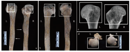
This study made an pathological analysis of an archaeological case example of human unilateral humeral hypoplasia from the Eastern Zhou Dynasty(770-221 BC) Guanzhuang site, located in Xingyang City, Henan Province, central China. Individual M45, a female (according to the morphology of the os coxa and skull, and long bones) aged approximately 30 years old at her time-of-death, presents severe abnormal morphological changes on the right humerus. From a macro perspective, abnormal shortening, flatter humeral head, higher anatomical neck, and lesser tubercle displaced anterior-distally were observed on the right humerus. Radiographic analysis showed that wider bone marrow cavity diameter and slight osteoporosis in the region of the deltoid tuberosity, and the cancellous bone at the trochanter of the right deltoid is more pronounced than on the left one, showing a honeycomb shape. In addition, bone defect was found at the region of the deltoid tuberosity and also below the anatomical neck. The asymmetric development of upper limb bones, including the humerus, is generally attributed to physiological or pathological induced bone remodeling. Physiological causes include genetic and behavioral factors. The asymmetry caused by physiological genetic factors does not affect the normal development of bone, and the degree of asymmetry is relatively low, in the range of 1% or less. Behavioral factors are mainly related to activities and bone loading patterns, including occupation, habitual activities, etc. The difference between two humerus caused by behavioral factors is reflected in the difference of the cross-section and the strength of the two humerus, most of which are transverse differences. Differential diagnosis was made in this study, including metabolic diseases, endocrine diseases, focal fibrocartilage hypoplasia, metaphyseal cartilage hypoplasia, Kashin Beck disease, osteogenesis imperfecta, humeral shortening and cartilage hypoplasia. According to the analysis, it is more likely that the humeral asymmetry found in M45 has affected the proximal growth centre of the right humerus, possibly linked to childbirth trauma, or the cessation of humeral growth and development caused by trauma to the growth plate in early childhood.

Key words: Eastern Zhou; humerus; development; pathology
| [1] | 夏洛特·罗伯茨. 疾病考古学(第三版)[M]. 济南: 山东画报出版社, 2010 |
| [2] | Amanda B. Bilateral Asymmetry of the Humerus During Growth and Development[J]. American Journal of Physical Antheropology, 2011, 145(4): 639-646 |
| [3] | Anna MK, Wioletta N, Antoine B, et al. Bilateral asymmetry of the humerus in Neandertals Australian aborigines and medieval humans[J]. American Journal of Physical Antheropology, 2018, 167(1): 46-60 |
| [4] | Steven EC, Vincenzo F. A Case of Marked Bilateral Asymmetry in the Upper Limbs of an Upper Palaeolithic Male from Barma Grande (Liguria), Italy[J]. International Journal of Osteoarchaeology, 1997, 7(1): 18-38 |
| [5] | Vladimír S, Margit B, Daniel S, et al. Human Manipulative Behavior in the Central European Late Eneolithic and Early Bronze Age: Humeral Bilateral Asymmetry[J]. American Journal of Physical Antheropology, 2007, 133(1): 669-681 |
| [6] | Kujanova M, Bigoni L, VelemIínska J, et al. Limb Bones Asymmetry and Stress in Medieval and Recent Populations of Central Europe[J]. International Journal of Osteoarchaeology, 2008, 18(5): 476-491 |
| [7] | Fields SJ, Spiers M, Hershkovitz I, et al. Reliability of reliability coefficients in the estimation of asymmetry[J]. American Journal of Physical Antheropology, 1995, 96(1): 83-87 |
| [8] | Sládek V, Berner M, Sosna D, et al. Human Manipulative Behavior in the Central European Late Eneolithic and Early Bronze Age: Humeral Bilateral Asymmetry[J]. American Journal of Physical Antheropology, 2007, 133: 669-681 |
| [9] | Kaya GZ, Adam DS, Christopher BR. Bilateral asymmetry and developmental plasticity of the humerus in modern humans[J]. American Journal of Physical Antheropology, 2021, 418-433 |
| [10] | Shaw CN, Stock JT. Habitual Throwing and Swimming Correspond With Upper Limb Diaphyseal Strength and Shape in Modern Human Athletes[J]. American Journal of Physical Antheropology, 2009, 140: 160-172 |
| [11] | 邓阳, 李宝华, 侯海峰, 等. 肱骨短小症致病相关基因及其变异的筛选与验证[J]. 中国地方病防治杂志, 2016, 31(9): 961-965 |
| [12] | 谢志勇, 谭为, 李旭. 儿童骨骺损伤的治疗及研究进展[J]. 中华实用儿科临床杂志, 2016, 31(11): 873-875 |
| [13] | 王建, 杨志明. 生长板损伤修复的研究进展[J]. 中国修复重建外科杂志, 2001, 5: 291-294 |
| [14] | Choi IH, Kim CJ, Cho TJ, et al. Focal fibrocartilaginous dysplasia of long bones: report of eight additional cases and literature review[J]. J Pediatr Orthop, 2000, 20(4): 421-427 |
| [15] | Chong JK, In HC, Tae JC, et al. The histological spectrum of subperiosteal fibrocartilaginous pseudotumor of long bone (focal fibrocartilaginous dysplasia)[J]. Pathology International, 1999, 49: 1000-1006 |
| [16] | Bell SN, Campbell PE, Cole WG, et al. Tibia vara caused by focal fibrocartilaginous dysplasia: three case reports[J]. J Bone Joint Surg Br, 1985, 67(5): 780-784 |
| [17] | 焦凤萍, 刘一志, 李宝华, 等. 肱骨短小症患者骨生长因子检测及意义[J]. 中国公共卫生, 2014, 30(9): 1184-1186 |
| [18] | Lieverse A, Metcalf M, Bazaliiskii V, et al. Pronounced Bilateral Asymmetry of the Complete Upper Extremity: A Case From the Early Neolithic Baikal, Siberia[J]. International Journal of Osteoarchaeology, 2008, 18: 219-239 |
| [19] | Kacki S, Duneufjardin P, Blanchard P, et al. Humerus Varus in a Subadult Skeleton from the Medieval Graveyard of LaMadeleine (Orléans, France)[J]. International Journal of Osteoarchaeology, 2013, 23(1): 119-126 |
| [20] | 惠夕平, 闫广宇, 王书成, 等. 河南荥阳官庄遗址周边考古调查简报[J]. 中国国家博物馆馆刊, 2017, 11: 22-31 |
| [21] | 邵象清. 人体测量手册[M]. 上海: 上海辞书出版社, 1985, 34-56 |
| [22] | 朱泓. 体质人类学[M]. 北京: 高等教育出版社, 2004, 92-106 |
| [23] | Zhou YW, Lu YP, He JN, et al. Bioarchaeological insights into disability: Skeletal dysplasia from the Iron Age northern China[J]. International Journal of Osteoarchaeology, 2021 |
| [24] | David W, Roberto M, Margherita M. A Case of Chondrodystrophic Dwarfism in the Italian Late Upper Paleolithic[J]. American Journal of Physical Anthropology, 1988, 75(4): 549-565 |
| [25] | 李崇, 富建华. 早产儿代谢性骨病诊治的研究进展[J]. 中国小儿急救医学, 2021, 28(1): 50-54 |
| [26] | 吕俊杰, 钟彩琴, 马长德, 等. 代谢性骨病患者BMD与TRAP、D-Pyr/Cr、NTX/Cr、HOP/Cr的检测及相关性研究[J]. 实用骨科杂志, 2016, 22(10): 902-905 |
| [27] | 张锦. 内分泌系统与疾病[M]. 上海: 上海科学技术出版社, 2008, 144-243 |
| [28] | 李新民, 蒋雯, 程克斌, 等. 儿童局灶性纤维软骨发育不良的影像特征[J]. 中国骨与关节杂志, 2017, 6(8): 582-586 |
| [29] | Jouve JL, Kohler R, Mubarak SJ, et al. Focal fibrocartilaginous dysplasia (“fibrous periosteal inclusion”): an additional series of eleven cases and literature review[J]. J Pediatr Orthop, 2007, 27(1): 75-84 |
| [30] | 赵定麟, 陈德玉, 赵杰. 现代骨科学[M]. 北京: 科学出版社, 2014, 50-58 |
| [31] | 中国疾病预防控制中心地方病控制中心网. 大骨节病[J]. 中国地方病防治杂志, 2019, 34(2): 211. |
| [32] | 李群伟, 次央, 侯海峰, 等. 肱骨短小症流行病学调查报告[J]. 中国预防医学杂志, 2012, 13(10): 721-723 |
| [33] | 李群伟, 龚弘强, 侯海峰, 等. 肱骨短小症患者临床体征与骨骼X线改变特征分析[J]. 中国地方病防治杂志, 2015, 30 (1): 4-6 |
| [34] | 刘明嫦, 马敬. 中国软骨发育不全特征分析及产前诊断方法探讨[J]. 云南医药, 2019, 40(2): 97-100 |
| [35] | 郑敏. 软骨发育不全 X 线诊断分析[J]. 中国伤残医学, 2014, 22(8): 194-195 |
| [36] | Stirland AJ. Asymmetry and activity-related change in the male humerus[J]. International Journal of Osteoarchaeology, 1993, 3(2): 105-113 |
| [37] | Schaeffer A. Spiral movement in man[J]. Journal of Morphology, 1928, 45(1): 293-399 |
| [38] | Eshed V, Gopher A, Galili E, et al. Musculoskeletal stress markers in Natufian Hunte-Gatherers and Neolothic Farmers in the Levant: The upper limb[J]. American Journal of Physical Anthropology, 2004, 123(4): 303-315 |
| [39] | Flachsmann R, Broom ND, Hardy AE, et al. Why is the adolescent joint particularly susceptible to osteochondral shear fracture?[J]. Clin Orthop Rel Res, 2000, 381: 212-21 |
| [40] | Micheli LJ. Pediatric and adolescent sports injury: recent trends[J]. In: Pandolf KB(Eds.). Exercise and sport science reviews[C]. New York: Macmillan, 1986, 359 |
| [41] | Salter RB, Harris WR. Injuries involving the epiphyseal plate[J]. J Bone Joint Surg, 1963, 45: 587-622 |
| [42] | 王香港, 万谦, 刘贺, 等. 组织工程软骨在生长板损伤修复治疗中的作用及特点[J]. 中国组织工程研究, 2021, 25(28): 4539-4545 |
| [43] | Beaty JH, Kasser JR. Rockwood and Wilkins' Fractures in Children[J]. J Bone Joint Surg, 2015, 88(10): 2313 |
| [44] | 洪意侠, 利春叶. 儿童骨骺损伤的诊断和治疗进展[J]. 罕少疾病杂志, 2021, 28(1): 107-110 |
/
| 〈 |
|
〉 |