

收稿日期: 2023-05-22
修回日期: 2023-07-20
网络出版日期: 2024-11-28
基金资助
中国科学院战略性先导科技专项(B类)(XDB26000000);国家自然科学基金(41872030)
A supernumerary tooth of the Liujiang Man from the Tongtianyan cave site in Guangxi
Received date: 2023-05-22
Revised date: 2023-07-20
Online published: 2024-11-28
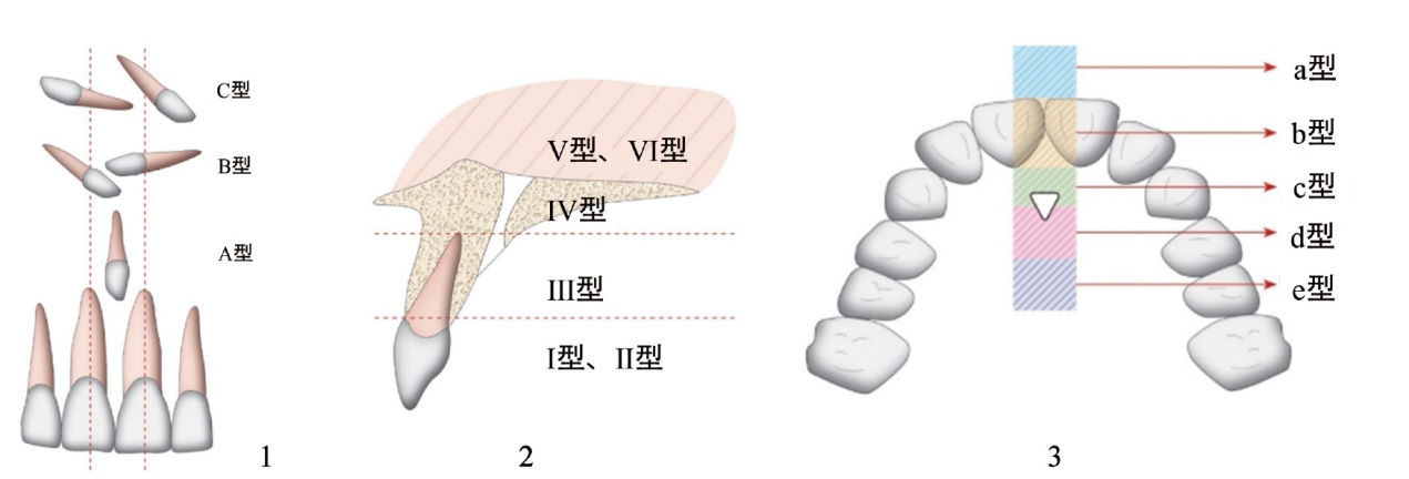
多生齿为正常齿列数目以外的牙齿,在现生人类中研究较多,但关于古人类多生齿的报道较少。晚更新世现代人-柳江人上颌硬腭中线右侧存在一颗圆锥型埋伏多生齿,通过高精度断层扫描和三维重建技术,发现其呈水平放置的状态,位置对应中腭区,齿尖朝向腭部后方,没有发现明显并发症。柳江人多生齿发生位置、形态、方向与包括全新世早期现代人在内的现生人类的部分案例重叠度较高,但是其综合特征具有特殊性。现代临床中有跟踪发现上颌前区的埋伏多生齿向腭部后方迁移的病例,因此推测柳江人多生齿特殊的发生状态可能是其迁移后的结果。柳江人作为东亚早期现代人的重要代表,不仅将中腭区水平放置多生齿的出现时间提早到更新世晚期,为埋伏多生齿的迁移现象提供了更早的化石证据,也为牙齿数目以及位置异常的古病理研究提供了新的参考案例。
孙琦雅慧 , 邢松 . 广西通天岩洞穴遗址柳江人的多生齿[J]. 人类学学报, 2024 , 43(06) : 1027 -1037 . DOI: 10.16359/j.1000-3193/AAS.2024.0012
Hyperdontia or supernumerary (or extra) teeth is one of the most common developmental anomalies in modern humans. Most supernumerary teeth are typically found in the maxillary anterior region, with about half occurring along the midline of the maxilla between the two central incisors, known as mesiodens. While supernumerary teeth have been extensively documented clinically, reports of such cases in human fossils are rare. This study describes a mesiodens embedded in the mid-palatal region of the Liujiang cranium, which is an early modern human from the Late Pleistocene in southern China discovered in the 1950s. The Liujiang mesiodens is partially exposed near the intermaxillary suture on the right side of the hard palate. High-resolution microcomputed tomography and three-dimensional virtual reconstruction further revealed that the mesiodens is conical in shape and horizontally positioned in the hard palate, with its long axis parallel to the intermaxillary suture. This mesiodens is situated distally to the long axis of the right central incisors with its cuspal apex facing the back of the palate, thus it is classified as a labiopalatinal type. Its crown corresponds to the position of the second premolar, away from the alveolar bone. Despite some distinctive features the location, shape, and direction of the Liujiang mesiodens overlapped with certain cases found in modern humans including that those from the Early Holocene. Based on clinical observations of pathological cases in modern humans, it is proposed that the Liujiang mesiodens may have migrated from the anterior region of the maxilla to the mid-palatal area. During this migration process, this supernumerary tooth adjusted its long axis from its original inverted form to its current horizontal position. This study provides evidence, dating back to the Late Pleistocene early modern humans for the occurrence of a horizontally positioned mesiodens in the mid-palatal region. Additionally, the Liujiang mesiodens presents early fossil evidence of supernumerary teeth in this special position and form than previously reported, as well as offers a case for the paleopathological study of abnormal number and position of teeth.
Key words: Early modern human; Supernumerary tooth; Paleopathology; Dental anomaly
| [1] | Rajab L, Hamdan M. Supernumerary teeth: review of the literature and a survey of 152 cases[J]. International Journal of Paediatric Dentistry, 2002, 12(4): 244-254 |
| [2] | Tyrologou S, Koch G, Kurol J. Location, complications and treatment of mesiodentes-a retrospective study in children[J]. Swed Dent J, 2005, 29(1): 1-9 |
| [3] | Garvey MT, Barry HJ, Blake M. Supernumerary teeth-an overview of classification, diagnosis and management[J]. Journal-Canadian Dental Association, 1999, 65(11): 612-616 |
| [4] | Mallineni SK. Supernumerary teeth: Review of the literature with recent updates[J]. In: Proceedings of the Conference Papers in Science[C]. Hindawi, 2014, 1-6 |
| [5] | Anthonappa R, King N, Rabie A. Diagnostic tools used to predict the prevalence of supernumerary teeth: a meta-analysis[J]. Dentomaxillofacial Radiology, 2012, 41(6): 444-449 |
| [6] | von Arx T. Anterior maxillary supernumerary teeth: A clinical and radiographic study[J]. Australian Dental Journal, 1992, 37(3): 189-195 |
| [7] | Liu Dg, Zhang Wl, Zhang Zy, et al. Three-dimensional evaluations of supernumerary teeth using cone-beam computed tomography for 487 cases[J]. Oral Surgery, Oral Medicine, Oral Pathology, Oral Radiology, and Endodontology, 2007, 103(3): 403-411 |
| [8] | Sedano HO, Gorlin RJ. Familial occurrence of mesiodens[J]. Oral Surgery, Oral Medicine, Oral Pathology, 1969, 27(3): 360-362 |
| [9] | Van Buggenhout G, Bailleul-Forestier I. Mesiodens[J]. European Journal of Medical Genetics, 2008, 51(2): 178-181 |
| [10] | Fernández Montenegro P, Valmaseda Castellón E, Berini Aytés L, et al. Retrospective study of 145 supernumerary teeth[J]. Medicina Oral, Patología Oraly Cirugía Bucal (Internet), 2006: 339-344 |
| [11] | Muhamad A, Moti M, Ornit C, et al. Histological and chemical analyses of mesiodens development and mineralization[J]. Archives of Oral Biology, 2018, 87: 191-195 |
| [12] | Henefer EP. Migrating mesiodens[J]. Oral Surgery, Oral Medicine, and Oral Pathology, 1967, 24(5): 636-637 |
| [13] | Asaumi J, Shibata Y, Yanagi Y, et al. Radiographic examination of mesiodens and their associated complications[J]. Dentomaxillofacial Radiology, 2004, 33(2): 125-127 |
| [14] | Shafi I, Gardner A, Brocklebank L. The migration of a mesiodens over eight years: a case report[J]. Orthodontic Update, 2011, 4(3): 89-92 |
| [15] | Kohli Gt, Verma P. Ectopic supernumerary tooth in the nasal cavity[J]. The Journal of Laryngology & Otology, 1970, 84(5): 537-538 |
| [16] | Ripamonti U, Petit JC, Thackeray JF. A supernumerary tooth in a 1.7 million-year-old Australopithecus robustus from Swartkrans, South Africa[J]. European Journal of Oral Sciences, 1999, 107(5): 317-321 |
| [17] | Sutton PR. Tooth eruption and migration theories: can they account for the presence of a 13,000-year-old mesiodens in the vault of the palate?[J]. Oral Surgery, Oral Medicine, Oral Pathology, 1985, 59(3): 252-255 |
| [18] | Villotte S, Ogden A, Trinkaus E. Dental abnormalities and oral pathology of the Pataud 1 upper paleolithic human[J]. Bulletins et Mémoires de la Société d'Anthropologie de Paris, 2018 |
| [19] | 吴汝康. 广西柳江发现的人类化石[J]. 古脊椎动物与古人类, 1959, 3(3): 97-104 |
| [20] | 吴汝康. 我国古人类学的新进展[J]. 科学通报, 1962, 8(8): 14-22 |
| [21] | 原思训, 陈铁梅, 高世君. 华南若干旧石器时代地点的铀系年代[J]. 人类学学报, 1986, 5(2): 179-190 |
| [22] | Shen G, Wang W, Wang Q, et al. U-Series dating of Liujiang hominid site in Guangxi, Southern China[J]. Journal of human evolution, 2002, 43(6): 817-829 |
| [23] | 王頠, 沈冠军, 周春林, 等. 柳江现代智人化石地点的地层及年代[J]. 第四纪研究, 2004, 24(3): 272-277 |
| [24] | 刘武, 吴秀杰, 汪良. 柳江人头骨形态特征及柳江人演化的一些问题[J]. 人类学学报, 2006, 25(3): 177-194 |
| [25] | 吴秀杰, 刘武, 董为, 等. 柳江人头骨化石的 CT 扫描与脑形态特征[J]. 科学通报, 2008, 53(13): 1570-1575 |
| [26] | 刘武, 吴秀杰, 李海军. 柳江人身体大小和形状——体重, 身体比例及相对脑量的分析[J]. 人类学学报, 2007, 4: 295-304 |
| [27] | 彼得, 布朗, 王谦, 等. 最初的蒙古人种吗?——对山顶洞 101 号、柳江及港川 Ⅰ号头骨的另一种看法[J]. 人类学学报, 1998, 17(4): 255-275 |
| [28] | 刘武, 曾祥龙. 第三臼齿退化及其在人类演化上的意义[J]. 人类学学报, 1996, 3: 185-199 |
| [29] | 于天平, 王荣. 恒牙列多生牙CBCT观测研究[J]. 影像研究与医学应用, 2022, 6(12): 132-134 |
| [30] | 章和平, 林凯烨, 蒋纯蓉, 等. 儿童上颌前部埋伏多生牙临床定位及拔除研究[J]. 临床口腔医学杂志, 2001, 2: 107-108+110 |
| [31] | Nagaveni N, Shashikiran N, Reddy VS. Surgical management of palatal placed, inverted, dilacerated and impacted mesiodens[J]. International Journal of Clinical Pediatric Dentistry, 2009, 2(1): 30-32 |
| [32] | Redwood C, Townsend G, Ghabriel M, et al. Under your nose: a rare finding during dissection provides insights into maxillary supernumerary teeth[J]. Australian Dental Journal, 2014, 59(3): 379-385 |
| [33] | Kim Y, Jeong T, Kim J, et al. Effects of mesiodens on adjacent permanent teeth: A retrospective study in Korean children based on cone-beam computed tomography[J]. International Journal of Paediatric Dentistry, 2018, 28(2): 161-169 |
| [34] | Goksel S, Agirgol E, Karabas HC, et al. Evaluation of prevalence and positions of mesiodens using cone-beam computed tomography[J]. Journal of Oral & Maxillofacial Research, 2018, 9(4): e1 |
| [35] | Toureno L, Park JH, Cederberg RA, et al. Identification of supernumerary teeth in 2D and 3D: Review of literature and a proposal[J]. Journal of Dental Education, 2013, 77(1): 43-50 |
| [36] | Kong J, Peng Z, Zhong T, et al. Clinical analysis of approach selection of extraction of maxillary embedded mesiodens in children[J]. Disease Markers, 2022, 2022: 6517024 |
| [37] | Xu GZ, Yang C, Yu CQ, et al. Embedded supernumerary teeth in the horizontal plate of palatine bone: report of 2 rare cases[J]. Journal of Oral and Maxillofacial Surgery, 2011, 69(5): 1295-1300 |
| [38] | Ma X, Jiang Y, Ge H, et al. Epidemiological, clinical, radiographic characterization of non-syndromic supernumerary teeth in Chinese children and adolescents[J]. Oral Diseases, 2021, 27(4): 981-992 |
| [39] | Zhao L, Liu S, Zhang R, et al. Analysis of the distribution of supernumerary teeth and the characteristics of mesiodens in Bengbu, China: a retrospective study[J]. Oral Radiology, 2021, 37(2): 218-223 |
| [40] | Tomczyk J, Szostek K, Lisowska-Gaczorek A, et al. A rare case of a supernumerary tooth (mesiodens) in an Iron Age (2470±35 BP) skeleton from Kozieg?owy (Poland)[J]. International Journal of Osteoarchaeology, 2020, 30(5): 736-742 |
| [41] | 吴新智. 周口店山顶洞人化石的研究[J]. 古脊椎动物与古人类, 1961, 3: 181-211 |
| [42] | Villa C, Davey J, Craig PJ, et al. The advantage of CT scans and 3D visualizations in the analysis of three child mummies from the Graeco-Roman Period[J]. Anthropol Anz, 2015, 72(1): 55-65 |
| [43] | Ives R. An unusual double supernumerary maxillary mesiodens in a Middle Iron Age skeleton from South Uist, Western Isles, Scotland[J]. Archives of oral biology, 2014, 59(6): 625-630 |
| [44] | 张全超. 北票喇嘛洞三燕文化墓地人骨的牙病[J]. 人类学学报, 2003, 22(1): 29-36 |
| [45] | Broehm CJ, Hunter LB, Boyd DK. A horizontal mesiodens in a child buried at Hank's site (41RB109), a prehistoric Plains Village Site in the Texas Panhandle[J]. Dental Anthropology Journal, 2011, 24(2-3): 55-58 |
| [46] | EM SB-O, Hurlen B, Humerfelt D. Changing positions of supernumerary teeth in the premaxilla: A radiographic study[J]. ASDC Journal of Dentistry for Children, 1985, 52(6): 428-430 |
| [47] | Hurlen B, Humerfelt D. Prevalence of premaxillary supernumerary teeth in Norwegian children: A radiographic study[J]. Dentomaxillofacial Radiology, 1984, 13(2): 109-115 |
| [48] | Sutton PR, Graze HR. The blood-vessel thrust theory of tooth eruption and migration[J]. Medical Hypotheses, 1985, 18(3): 289-295 |
| [49] | Bjork A. Postnatal growth and development of the maxillary complex[J]. Factors Affecting the Growth of the Midface, 1976: 61-69 |
| [50] | 彭兆伟, 钟天航, 韩剑丽, 等. 173例儿童上颌前部埋伏多生牙的回顾性研究[J]. 北京口腔医学, 2020, 28(1): 37-40 |
| [51] | Anthonappa R, King N, Rabie A. Aetiology of supernumerary teeth: a literature review[J]. European Archives of Paediatric Dentistry, 2013, 14(5): 279-288 |
| [52] | Tiwari M, Jaiswal S, Mitra GV, et al. Reversely paced mesiodens in mid-palatal Raphe Area: a rare case report[J]. International Journal of Innovative Research and Advanced Studies (IJIRAS), 4(2): 303-306 |
| [53] | Avsever H, Gunduz K, Orhan K, et al. An inverted eruption of mesiodens: Report of a rare case[J]. Clinical and Experimental Health Sciences, 2012, 2(1): 37-39 |
| [54] | Chopra S, Joshi M. Mesiodens erupted in the nasal cavity: report of a case[J]. Oral Surgery, Oral Medicine, Oral Pathology, 1969, 28(6): 856-858 |
/
| 〈 |
|
〉 |
 京ICP证05002819号-3