

颅骨板障静脉的三维复原及其在人类演化中的意义
收稿日期: 2025-04-11
录用日期: 2025-06-06
网络出版日期: 2025-08-07
基金资助
国家重点研发计划(2023YFF0804502);国家自然科学基金(42372001);国家自然科学基金(42472006)
Three dimensional reconstruction of cranial diploic veins and its significance in human evolution
Received date: 2025-04-11
Accepted date: 2025-06-06
Online published: 2025-08-07
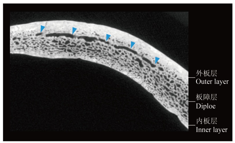
板障静脉是颅骨骨壁内部的复杂血管网络,在颅脑血液循环中发挥重要作用。自18世纪初发现该结构以来,受限于传统的解剖学观测方法,发表的相关研究成果非常匮乏,其形态变异与功能特征存在较多疑问。近年来,借助CT扫描设备和新开发的三维建模复原技术,板障静脉的研究正在突破瓶颈限制。新研究注意到板障静脉的传统分类命名缺少广泛适用性,因此重新确认了板障静脉的分布特点,并发现了其在脑脊液循环以及大脑冷却等方面的功能和意义。于古人类化石研究而言,则进一步注意到了板障静脉在不同种属间的差异,该研究有助于古人类化石的分类学讨论。此外,板障静脉可能参与了人类大脑演化,与颅容量以及大脑局部形状存在关联。本文在梳理板障静脉的研究历史和最新的研究进展的基础上,总结了其技术难点及未来的研究方向,以期促进国内相关研究的开展。
惠家明 . 颅骨板障静脉的三维复原及其在人类演化中的意义[J]. 人类学学报, 2025 , 44(04) : 545 -555 . DOI: 10.16359/j.1000-3193/AAS.2025.0059
Diploic veins are a complex vascular network within the cranial wall, playing a significant role in human cranial blood circulation. Since their discovery in the early 18th century, research on this structure has been limited by the traditional dissection methods, leaving many questions unresolved regarding their morphological variations, functions, and implications for human evolution. Recent advances in CT and MR imaging and newly-developed 3D reconstruction techniques have overcome previous bottlenecks, which can detect the morphological details of these vascular structures. New studies have revealed that traditional classifications of diploic veins lack broad applicability, while also redefining their distribution areas and patterns and identifying functional roles in cerebrospinal fluid circulation and cerebral thermoregulation, which may be essential to human brain physiology. Research on hominin fossils has further highlighted interspecific differences in diploic veins, offering potential applications in future taxonomical discussions. The current evidence indicates that the diploic vascular pattern can identify Neanderthal cranial fossils from other Homo species, and it also help differentiate non-human primates from hominins. Additionally, diploic veins may have contributed to human brain evolution, with possible links to brain volume and regional endocast morphology. To clarify the mechanism behind the variation and evolution, further studies are warranted to investigate the diploic vascular morphology and physiology in large extant human populations and also in hominin fossils. This paper reviews the history of diploic vein research and synthesizes recent advancements, summarizing methodologies, technical challenges, and future directions to promote further studies in this field.
Key words: hominins; Micro-CT; 3D reconstruction; brain evolution; taxonomy
| [1] | Hui J. Comparative anatomy of the diploic vessels in hominids: implications for the evolutionary history of human cranial blood circulation[D]. Paris: Sorbonne University, 2024 |
| [2] | García-gonzález U, Cavalcanti DD, Agrawal A, et al. The diploic venous system: surgical anatomy and neurosurgical implications[J]. Neurosurgical Focus, 2009, 27(5): E2 |
| [3] | Pí?ová H, Rangel De Lázaro G, Velemínsky P, et al. Craniovascular traits in anthropology and evolution: from bones to vessels[J]. Journal of Anthropological Sciences, 2017, 95: 35-65 |
| [4] | Bruner E, Mantini S, Musso F, et al. The evolution of the meningeal vascular system in the human genus: From brain shape to thermoregulation[J]. American Journal of Human Biology, 2011, 23(1): 35-43 |
| [5] | Falk D. Evolution of cranial blood drainage in hominids: Enlarged occipital/marginal sinuses and emissary foramina[J]. American Journal of Physical Anthropology, 1986, 70(3): 311-324 |
| [6] | Hui J, Balzeau A. The diploic venous system in Homo neanderthalensis and fossil Homo sapiens: A study using high-resolution computed tomography[J]. American Journal of Biological Anthropology, 2023, 182(3): 412-427 |
| [7] | Grimaud-hervé D. Endocranial Vasculature[M]. In: Schwartz JH, Tattersall I, Holloway RL(eds.). The human fossil record, brain endocasts - The paleoneurological evidence: volume 3. New York: Wiley-Liss, 2002: 273-277 |
| [8] | Breschet G. Recherches anatomiques, physiologiques et pathologiques sur le système veineux[M]. Paris: Villeret, 1829 |
| [9] | Hershkovitz I, Greenwald C, Rothschild BM, et al. The elusive diploic veins: Anthropological and anatomical perspective[J]. American Journal of Physical Anthropology, 1999, 108(3): 345-358 |
| [10] | Schünke M, Schulte E, Schumacher U, et al. Tête, cou et neuro-anatomie[M]. Louvain-la-Neuve: De Boeck supérieur, 2016 |
| [11] | Tsutsumi S, Ono H, Ishii H, et al. Diploic veins of the cranial base: an anatomical study using magnetic resonance imaging[J]. Surgical and Radiologic Anatomy, 2019, 41(9): 1029-1036 |
| [12] | Lachkar S, Dols M M, Ishak B, et al. The diploic veins: A comprehensive review with clinical applications[J]. Cureus, 2019, 11(4): e4422 |
| [13] | Tsutsumi S, Nakamura M, Tabuchi T, et al. Calvarial diploic venous channels: an anatomic study using high-resolution magnetic resonance imaging[J]. Surgical and Radiologic Anatomy, 2013, 35(10): 935-941 |
| [14] | Yamashiro K, Muto J, Wakako A, et al. Diploic veins as collateral venous pathways in patients with dural venous sinus invasion by meningiomas[J]. Acta Neurochirurgica, 2021, 163(6): 1687-1696 |
| [15] | Tsutsumi S, Ogino I, Miyajima M, et al. Cranial arachnoid protrusions and contiguous diploic veins in CSF drainage[J]. American Journal of Neuroradiology, 2014, 35(9): 1735-1739 |
| [16] | Tsutsumi S, Ogino I, Miyajima M, et al. Cerebrospinal fluid drainage through the diploic and spinal epidural veins[J]. Journal of Anatomy, 2015, 227(3): 297-301 |
| [17] | Falk D. Brain evolution in Homo: The “radiator” theory[J]. Behavioral and Brain Sciences, 1990, 13(2): 333-344 |
| [18] | Poblet E, Jimenez F, Escario-Travesedo E, et al. Eccrine sweat glands associate with the human hair follicle within a defined compartment of dermal white adipose tissue[J]. British Journal of Dermatology, 2018, 178(5): 1163-1172 |
| [19] | Bruner E, Eisova S. Vascular microforamina and endocranial surface: Normal variation and distribution in adult humans[J]. The Anatomical Record, 2024 |
| [20] | Rangel De Lázaro G, Neubauer S, Gunz P, et al. Ontogenetic changes of diploic channels in modern humans[J]. American Journal of Physical Anthropology, 2020, 173(1): 96-111 |
| [21] | Rangel De Lázaro G,De La Cuétara JM,Pí?ová H, et al. Diploic vessels and computed tomography: Segmentation and comparison in modern humans and fossil hominids[J]. American Journal of Physical Anthropology, 2016, 159(2): 313-324 |
| [22] | Yamashiro K, Wakako A, Omi T, et al. Evaluating diploic vein blood flow using time-resolved whole-head computed tomography angiography and determining the positional relationship between typical craniotomy approaches and diploic veins in patients with meningioma[J]. Acta Neurochirurgica, 2022, 164(11): 2999-3010 |
| [23] | Tsutsumi S, Ono H, Ishii H. Correlation of the external occipital protuberance with venous sinuses: A magnetic resonance imaging study[J]. Surgical and Radiologic Anatomy, 2022, 44(7): 999-1006 |
| [24] | Eisová S, Menéndez LP, Velemínsky P, et al. Craniovascular variation in four late Holocene human samples from southern South America[J]. The Anatomical Record, 2023, 306(1): 143-161 |
| [25] | Masters BR. Fractal analysis of the vascular tree in the human retina[J]. Annual Review of Biomedical Engineering, 2004, 6(1): 427-452 |
| [26] | Sanghera B, Banerjee D, Khan A, et al. Reproducibility of 2D and 3D fractal analysis techniques for the assessment of spatial heterogeneity of regional blood flow in rectal cancer[J]. Radiology, 2012, 263(3): 865-873 |
| [27] | Di Ieva A. The fractal geometry of the brain[M]. New York: Springer, 2016 |
| [28] | Mandelbrot BB. The fractal geometry of nature[M]. Oxford: Freeman, 1982 |
| [29] | Liu W, Athreya S, Xing S, et al. Hominin evolution and diversity: a comparison of earlier-Middle and later-Middle Pleistocene hominin fossil variation in China[J]. Philosophical Transactions of the Royal Society B: Biological Sciences, 2022, 377(1847): 20210040 |
| [30] | Roksandic M, Radovi? P, Wu X, et al. Resolving the “muddle in the middle”: The case for Homo bodoensis sp. nov.[J]. Evolutionary Anthropology: Issues, News, and Reviews, 2022, 31(1): 20-29 |
| [31] | Wu X, Pei S, Cai Y, et al. Morphological and morphometric analyses of a late Middle Pleistocene hominin mandible from Hualongdong, China[J]. Journal of Human Evolution, 2023, 182: 103411 |
| [32] | Wu X, Bruner E. The endocranial anatomy of Maba 1[J]. American Journal of Physical Anthropology, 2016, 160(4): 633-643 |
| [33] | 吴秀杰. 中更新世晚期许家窑人化石的研究进展[J]. 人类学学报, 2024, 43(1): 5-18 |
| [34] | 刘武, 吴秀杰, 邢松. 更新世中期中国古人类演化区域连续性与多样性的化石证据[J]. 人类学学报, 2019, 38(4): 473-490 |
| [35] | Balzeau A, Albessard-Ball L, Kubicka AM, et al. Frontal sinuses and human evolution[J]. Science Advances, 2022, 8(42): eabp9767 |
| [36] | Balzeau A, Albessard-Ball L, Kubicka AM, et al. Frontal sinus variation in extant species of the genera Pan, Gorilla and Homo[J]. Bulletins et mémoires de la société d’anthropologie de Paris, 2021, 33(2): 27-52 |
| [37] | Balzeau A, Buck LT, Albessard L, et al. The Internal Cranial Anatomy of the Middle Pleistocene Broken Hill 1 Cranium[J]. PaleoAnthropology, 2017: 107-138 |
| [38] | Quam R, Martínez I, Rosa M, et al. Early hominin auditory capacities[J]. Science Advances, 2015, 1(8): e1500355 |
| [39] | Conde-Valverde M, Martínez I, Quam R, et al. The ear of the Sima de los Huesos hominins (Atapuerca, Spain)[J]. The Anatomical Record, 2024, 307(7): 2410-2424 |
| [40] | Wu X, Crevecoeur I, Liu W, et al. Temporal labyrinths of eastern Eurasian Pleistocene humans[J]. Proceedings of the National Academy of Sciences, 2014, 111(29): 10509-10513 |
| [41] | Li ZY, Wu X, Zhou LP, et al. Late Pleistocene archaic human crania from Xuchang, China[J]. Science, 2017, 355(6328): 969-972 |
| [42] | Saban R. Image of the human fossil brain: Endocranial casts and meningeal vessels in young and adult subjects[M]. In: Origins of the human brain. Oxford: Oxford University Press, 1995: 11-39 |
| [43] | Kunz AR, Iliadis C. Hominid evolution of the arteriovenous system through the cranial base and its relevance for craniosynostosis[J]. Child’s Nervous System, 2007, 23(12): 1367-1377 |
| [44] | Bruner E, Sherkat S. The middle meningeal artery: from clinics to fossils[J]. Child’s Nervous System, 2008, 24(11): 1289-1298 |
| [45] | Bruner E, Mantini S, Perna A, et al. Fractal dimension of the middle meningeal vessels: variation and evolution in Homo erectus, Neanderthals, and modern humans[J]. European Journal of Morphology, 2006, 42(4-5): 217-224 |
| [46] | Bruner E, De La Cuétara JM, Masters M, et al. Functional craniology and brain evolution: from paleontology to biomedicine[J]. Frontiers in Neuroanatomy, 2014, 8: 19 |
| [47] | Juskys R, Rocka S, Suchomlinov A. Anatomical variations of superior sagittal sinus and tributary bridging veins: A cadaveric study[J]. Cureus, 2022, 14(2):e21979 |
| [48] | Anzelmo M, Ventrice F, Barbeito-Andrés J, et al. Ontogenetic changes in cranial vault thickness in a modern sample of Homo sapiens[J]. American Journal of Human Biology, 2015, 27(4): 475-485 |
| [49] | Balzeau A. Thickened cranial vault and parasagittal keeling: Correlated traits and autapomorphies of Homo erectus?[J]. Journal of Human Evolution, 2013, 64(6): 631-644 |
| [50] | Hui J, Balzeau A. Investigating the relationship between cranial bone thickness and diploic channels: A first comparison between fossil Homo sapiens and Homo neanderthalensis[J]. The Anatomical Record, 2024, 307(6): 2036-2046 |
/
| 〈 |
|
〉 |
 京ICP证05002819号-3