

主管:中国科学院
主办:中国科学院古脊椎动物与古人类研究所
出版:科学出版社
主办:中国科学院古脊椎动物与古人类研究所
出版:科学出版社
人类学学报 ›› 2026, Vol. 45 ›› Issue (04): 695-708.doi: 10.16359/j.1000-3193/AAS.2025.0079cstr: 32091.14.j.1000-3193/AAS.2025.0079
王师慧1( ), 魏东2(), 党志豪3
收稿日期:2024-07-12
修回日期:2025-04-24
出版日期:2026-08-15
发布日期:2026-08-12
通讯作者:
魏东,博士,主要从事考古人类学研究。E-mail: 作者简介:王师慧,博士研究生,主要从事人类骨骼考古学研究。E-mail: wangsh22@jlu.edu.cn
基金资助:
WANG Shihui1(), WEI Dong2(), DANG Zhihao3
Received:2024-07-12
Revised:2025-04-24
Online:2026-08-15
Published:2026-08-12
摘要:
2023年,在新疆库车友谊路墓地的发掘过程中,于WM341墓主左侧胫骨发现溶骨性损伤。该个体为14~20岁的青年男性个体。损伤具体表现为左侧胫骨骨干有两处明显的虫蚀状骨损痕迹;左侧腓骨、右侧胫腓骨未发现病理现象;其余部位骨骼除部分胸椎发生黄韧带骨化之外,均未见异常。应用正置显微镜、X光和CT断层扫描等技术手段对病灶做进一步观察和分析后,结合现代临床案例研究,从流行病学和病理表现两个方面排除了急性血源性骨髓炎、低级别中央型骨肉瘤、孤立性骨囊肿、朗格汉斯细胞组织细胞增生症、恶性骨巨细胞瘤等发生的可能性。该溶骨性损伤是尤文氏肉瘤的可能性最大。尤文氏肉瘤恶性程度高,或为该个体死亡的直接死因。该案例为目前国内古代样品所见尤文氏肉瘤的首次报道。
中图分类号:
王师慧, 魏东, 党志豪. 新疆库车友谊路墓地居民溶骨性损伤的古病理学[J]. 人类学学报, 2026, 45(04): 695-708.
WANG Shihui, WEI Dong, DANG Zhihao. Paleopathology of a person with osteolytic injury from the Youyilu Cemetery, Kuqa, Xinjiang[J]. Acta Anthropologica Sinica, 2026, 45(04): 695-708.
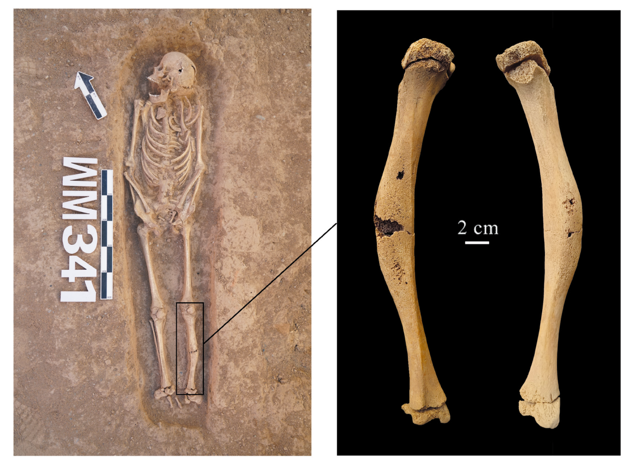
图1 WM341考古发掘现场及患病骨骼详细情况
Fig.1 Excavation and infected tibia of the individual in WM341
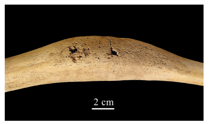
图2 患病骨骼细部情况
Fig.2 Details on the infected bone
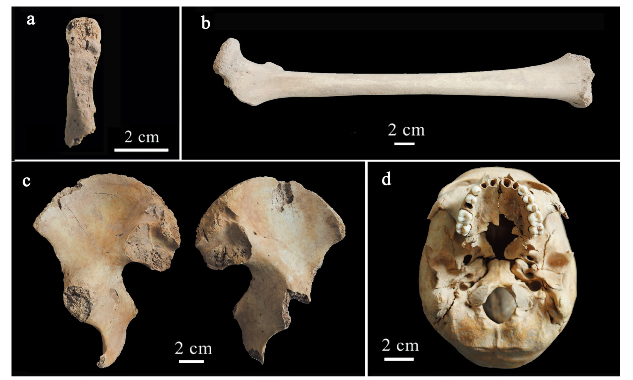
图3 WM341骨骼发育情况 a.左侧耻骨联合面Left pubic symphysis;b.右侧股骨Right femur;c.髋骨Os coxae;d.颅底Base of skull
Fig.3 Skeletal development of the individual in WM341
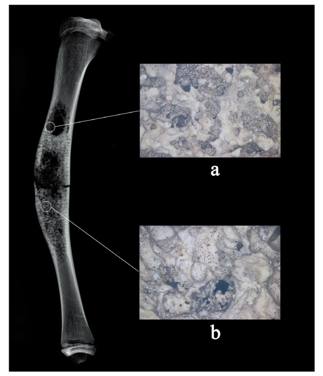
图4 正置显微镜下的病灶区域虫蚀状痕迹(50×)
Fig.4 Moth-eaten damages over the osteolytic lesion under the upright microscope 表面痕迹Osteolytic damage on the surface: a. 2号病灶 second lesion; b. 1号病灶first lesion
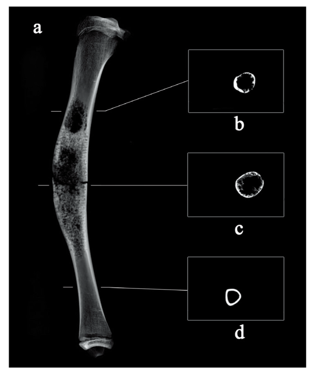
图5 左侧胫骨X光侧位片及CT断层影像 a.左侧胫骨left tibia; b. 2号病灶second lesion; c. 1号病灶first lesion; d. 未受损区域segment free from damage
Fig.5 X-ray and CT imaging over the osteolytic lesion of left tibia
| [1] | Ortner DJ. Identification of Pathological Conditions in Human Skeletal Remains[M]. Orlando: Elsevier Science, 2003, 503-504 |
| [2] | 邵象清. 人体测量手册[M]. 上海: 上海辞书出版社, 1985, 34-56 |
| [3] | 朱泓. 体质人类学[M]. 北京: 高等教育出版社, 2004, 92-106 |
| [4] | Buikstra JE, Ubelaker DH. Standards for data collection from human skeletal remains[M]. Fayetteville: Arkansas Archaeological Survey, 1994, 15-38 |
| [5] | 高津光洋. 法医尸检手册[M]. 北京: 人民卫生出版社, 2021, 303-327+364-367 |
| [6] | Krogman W, Iscan M. The Human Skeleton in Forensic Medicine[M]. Springfield: Charles C, Thomas, 1986, 16-43 |
| [7] | McKern T, Stewart T. Skeletal Age Changes in Young American Males, Analyzed from the Standpoint of Identification[A]. In: Technical Report EP-45[C]. Natick: Headquarters, Quartermaster Research and Development Command, 1957, 25-42 |
| [8] | Redfiled A. A New Aid to Aging Immature Skeletons: Development of the Occipital Bone[J]. American Journal of Physical Anthropology, 1970, 33: 217-220 |
| [9] | Suchey J, Owings P, Wiseley D, et al. Skeletal Aging of Unidentified Persons[A]. In: Ted A, Jane E. Human Identification: Case Studies in Forensic Anthropology[C]. Springfield: Charles C, Thomas, 1984, 278-297 |
| [10] | Ubelaker D. Human Skeletal Remains[M]. Washington DC: Taraxacum Press, 1989, 29-65 |
| [11] | Ubelaker D. The Estimation of Age at Death from Immature Human Bone[A]. In: Iscan M. Age Markers in the Human Skeleton[C]. Springfield: Charles C. Thomas, 1989, 55-70 |
| [12] |
Dion E, Gmef C, Miquel A, et al. Bone involvement in erdheim-ehester disease: imaging findings including periostitis and partial epiphyseal involvement[J]. Radiology, 2006, 238(2): 632-639
doi: 10.1148/radiol.2382041525 URL |
| [13] | De Filippo M, Ingegnoli A, Carloni A, et al. Erdheim-Chester disease: clinical and radiological findings[J]. Radiologica Medica, 2009, 114(8): 1319-1329 |
| [14] | Kempson RL. Ossifying fibroma of long bones: a light and electron microscopic study[J]. Archives of Pathology, 1996, 82: 218-223 |
| [15] |
Campanacci M, Laus M. Osteofibrous dysplasia of the tibia and fibula[J]. Bone and Joint Surgery, 1981, 63(3): 367-375
doi: 10.2106/00004623-198163030-00007 URL |
| [16] |
Kyle RA, Gertz MA, Witzig TE, et al. Review of 1027 patients with newly diagnosed multiple myeloma[J]. Mayo Clinic Proceedings, 2003, 78(1): 21-33
doi: 10.4065/78.1.21 pmid: 12528874 |
| [17] | 胡聪, 匡春梅, 周文. 多发性骨髓瘤骨病发病机制的研究进展[J]. 中南大学学报(医学版), 2023, 48(9): 1403-1410 |
| [18] | 胥少汀, 葛宝丰, 徐印坎. 实用骨科学[M]. 第4版, 北京: 人民军医出版社, 2012, 1751 |
| [19] | 余建国, 姚安晋. 四肢长骨骨干结核的X线诊断(附9例报告)[J]. 实用放射学杂志, 2000, 6: 372-373 |
| [20] |
Sudhir KG, Akash S, Anubhav M. Primary tuberculosis of the fibular diaphysis: A rare case report[J]. International Journal of Surgery Case Reports, 2020, 74: 140-143
doi: S2210-2612(20)30618-0 pmid: 32836210 |
| [21] | Mahmood M, Nassiri MC, Mohamed O, et al. Diaphyseal tuberculosis of tibia- rare occurrence: case report and review of literature[J]. Bone Reports. Recomment, 2016, 2 |
| [22] | Hsieh C, Miltner LE, Chang C. Tuberculosis of the shaft of the large long bones of the extremities[J]. Bone and Joint Surgery, 1934, 16: 545-563 |
| [23] | Ewing J. Diffuse endothelioma of bone[J]. Proceedings of the New York Pathological Society, 1921, 21(1): 17-24 |
| [24] |
Sanker S, Lessnick SL. Promiscuous partnerships in Ewing’s sarcoma[J]. Cancer Genet, 2011, 204: 351-365
doi: 10.1016/j.cancergen.2011.07.008 URL |
| [25] | Novik VI, Krasil LA, Kolygin BA, et al. Cytological diagnosis and prognosis in Ewing’s sarcoma in children[J]. Archives of Pathology, 2005, 67(2): 22-25 |
| [26] |
Von Eisenhart R, Toepfer A, Salzmann M, et al. Primary malignant bone tumors[J]. Orthopade, 2011, 40(12): 1121-1142
doi: 10.1007/s00132-011-1866-7 pmid: 22130624 |
| [27] | 李洪涛, 李董董, 杨晓笛, 等. 尤文氏肉瘤误诊为非骨化性纤维瘤1例[J]. 中国骨伤, 2017, 30(1): 68-70 |
| [28] | 王强修, 孙成良, 张光辉. 骨肿瘤诊断与治疗[M]. 北京: 中国医药科技出版社, 2010, 136 |
| [29] | 童成文, 陈光斌, 胡久民, 等. 原发性颅骨尤文氏肉瘤1例[J]. 医学影像学杂志, 2020, 30(4): 648+685 |
| [30] | 孙辉, 李邦国, 黄婷婷, 等. 原发性肋骨尤文氏肉瘤一例[J]. 临床放射学杂志, 2016, 35(5): 802-803 |
| [31] | 刘俊, 张文君, 蒋秋华. 腰椎管内原发性尤文氏肉瘤/原始神经外胚层肿瘤1例[J]. 中国临床神经外科杂志, 2023, 28(4): 287-288 |
| [32] | 吴恩惠, 张景荣, 曹来宾, 等. 放射学[M]. 北京: 人民卫生出版社, 1996, 182-184 |
| [33] | Resnick D. Diagnosis of bone and joint disorders(4th edition)[M]. Philadelphia: WB Saunders Co, 2002, 4060 |
| [34] | 张本霖, 张金祥. 尤文肉瘤早期误诊为化脓性骨髓炎1例[J]. 临床军医杂志, 2007, 8(5): 699 |
| [35] | 尧浩, 杨宜, 庞李贺, 等. 锁骨尤文氏肉瘤锁骨全切术:1例报告与综述[J]. 中国矫形外科杂志, 2021, 29(23): 2154-2156+2161 |
| [36] | Gibbs CP, Weber K, Scar borough MT, et al. Malignant bone tumors[J]. Instructional Course Lectures, 2008, 51: 413-428 |
| [37] | 田竞, 祖启明, 项良碧. 化脓性骨髓炎[J]. 中国实用乡村医生杂志, 2006, 8: 16-17 |
| [38] |
Riise OR, Kirkhus E, Handeland KS, et al. Childhood osteomyelitis-incidence and differentiation from other acute onset musculoskeletal features in a population-based study[J]. BMC Pediatrics, 2008, 8(1): 45-51
doi: 10.1186/1471-2431-8-45 URL |
| [39] | 胡金平. 浅析急性化脓性骨髓炎的影像诊断[J]. 当代医学, 2010, 16(15): 53-54 |
| [40] | Song KM, Sloboda JF. Acute hematogenous osteomyelitis in children[J]. The Journal of the American Academy of Orthopaedic Surgery, 2001, 9(3): 166-175 |
| [41] | 陈孝平, 汪建平, 赵继宗. 外科学[M]. 第9版, 北京: 人民卫生出版社, 2018, 738-739 |
| [42] | 刘峰. MRI在急性化脓性骨髓炎早期诊断中的应用探究[J]. 现代医用影像学, 2017, 26(2): 377-379 |
| [43] | 吴孟超, 吴在德, 黄家驷. 外科学[M]. 第7版, 北京: 人民卫生出版社, 2008, 2982-3004 |
| [44] | 胥少汀, 葛宝丰, 徐印坎. 实用骨科学[M]. 第4版, 北京: 人民军医出版社, 2012, 1557 |
| [45] | 李继承, 曾圆山, 周莉, 等. 组织学与胚胎学[M]. 第9版, 北京: 人民卫生出版社, 2018, 29 |
| [46] | 陈孝平, 汪建平, 赵继宗. 外科学[M]. 第9版, 北京: 人民卫生出版社, 2018, 776-777 |
| [47] |
Unni KK, Dahlin DC, Mc Leod RA, et al. Intraosseous well-differentiated osteosarcoma[J]. Cancer, 1977, 40(3): 1337-1347
pmid: 269000 |
| [48] | Unni KK. Bone Tumors[M]. New York: Churchill Livingstone, 1988, 11: 107-133 |
| [49] | 魏清柱. WHO骨肿瘤分类[J]. 第5版,诊断病理学杂志, 2021, 28(6): 497-498 |
| [50] | Fletcher CD, Unni KK, Mertens F. World Health Organization classification of tumors: Pathology & genetics tumours of soft tissue and bone[M]. Lyon: IARC Press, 2002, 275-276 |
| [51] | 巴得热力·特尔巴图, 袁治国, 秦进, 等. 低度恶性中央型骨肉瘤的研究进展[J]. 甘肃科技, 2022, 38(21): 130-133 |
| [52] | 赵成. 低度恶性中央性骨肉瘤(LGCOS)的临床诊治分析[D]. 硕士研究生毕业论文, 济南: 山东大学, 2014, 36 |
| [53] |
Kurt A, Unni K, Mc Leod R, et al. Low-grade intraosseous osteosarcoma[J]. Cancer, 1990, 65(6): 1418-1445
doi: 10.1002/1097-0142(19900315)65:6<1418::aid-cncr2820650629>3.0.co;2-q pmid: 2306687 |
| [54] |
Unni K. Case report 136. Central low-grade osteosarcoma of tibia[J]. Skeletal Radiology, 1981, 6(1): 65-71
pmid: 6936837 |
| [55] |
Fukunaga M. Low-grade central osteosarcoma of the skull[J]. Pathology, Research and Practice, 2005, 201(2): 131-135
pmid: 15901134 |
| [56] |
Diniz A, Alencar R, Garcia R, et al. Low-grade central osteosarcoma of the mandible: a case study report[J]. Oral Surgery, Oral Medicine, Oral Pathology, Oral Radiology, and Endodontology, 2007, 103(2): 246-252
doi: 10.1016/j.tripleo.2005.11.006 URL |
| [57] |
Bugnone AN, Temple HT, Pitcher JD. Low-grade central osteosarcoma of the foot and ankle: radiographic and pathologic features in two patients: case report and literature review[J]. Foot Ankle International, 2005, 26(6): 494-500
doi: 10.1177/107110070502600612 URL |
| [58] | 易申德, 吴欣乐, 杨成茂, 等. 孤立性骨囊肿治疗的对比研究[J]. 实用医学杂志, 2017, 33(16): 2732-2735 |
| [59] | Jaffe HL, Lichtenstein L. Non-osteogenic fibroma of bone[J]. The American Journal of Pathology, 1942, 18(2): 205-221 |
| [60] | 荣独山. X线诊断学[M]. 第2版, 上海: 上海科学技术出版社, 2000, 280-281 |
| [61] | 王强修, 孙成良, 张光辉. 骨肿瘤诊断与治疗[M]. 北京: 中国医药科技出版社, 2010, 125-126 |
| [62] | 胥少汀, 葛宝丰, 徐印坎. 实用骨科学[M]. 第4版, 北京: 人民军医出版社, 2012, 1785 |
| [63] | 俎战露, 张洲, 郝晓东. 多发性孤立性骨囊肿1例[J]. 西北国防医学杂志, 2006, 6: 431 |
| [64] | 陈孝平, 汪建平, 赵继宗. 外科学[M]. 第9版, 北京: 人民卫生出版社, 2018, 779 |
| [65] | Lichtenstein L. Histocytosis: integration of eosinophilic granuloma of bone, Letterer-Siwe disease, and Schüller-Christian disease as related manifestations of a single nosologic entity[J]. Archives of Pathology, 1953, 56(1): 84-102 |
| [66] | 何平根, 侯婕妤, 王广平. 儿童局灶性朗格汉斯细胞组织细胞增生症一例[J]. 实用医技杂志, 2020, 27(2): 272-273 |
| [67] | 胥少汀, 葛宝丰, 徐印坎. 实用骨科学[M]. 第4版, 北京: 人民军医出版社, 2012, 1800 |
| [68] | 王强修, 孙成良, 张光辉. 骨肿瘤诊断与治疗[M]. 北京: 中国医药科技出版社, 2010, 205 |
| [69] |
Beytemür O, Tetikkurt S, Albay C, et al. Liposclerosing myxofibrous tumor: a rare tumor of proximal femur[J]. Eklem Hastalik Cerrahisi, 2017, 28(3): 210-213
doi: 10.5606/ehc.2017.48394 URL |
| [70] |
Hoover KB, Rosenthal DI, Mankin H. Langerhans cell histiocytosis[J]. Skeletal Radiology, 2007, 36(2): 95-104
doi: 10.1007/s00256-006-0193-2 pmid: 17028900 |
| [71] | WHO classification of tumours editorial board. Soft tissue and bone tumours, WHO classification of tumours (5th edition)[M]. Lyon: IARC Press, 2020, 321-525 |
| [72] | 赵建明, 杨军舰, 宋传涛, 等. 长骨朗格汉斯细胞组织细胞增生症的影像诊断[J]. 医学影像学杂志, 2023, 33(5): 840-842+846 |
| [73] |
Rosenberg AE. WHO Classification of Soft Tissue and Bone, fourth edition: summary and commentary[J]. Current Opinion in Oncology, 2013, 25(5): 571-573
doi: 10.1097/01.cco.0000432522.16734.2d pmid: 23942303 |
| [74] | Gamberi G, Serra M, Ragazzini P, et al. Indentifications of markers of possible prognostic value in 57 giant cell tumors of bone[J]. Oncology Reports, 2003, 10(2): 351-356 |
| [75] |
Campanacci M, Baldini N, Boriani S, et al. Giant-cell tumor of bone[J]. Bone and Joint Surgery, 1987, 69(1): 106-114
doi: 10.2106/00004623-198769010-00018 URL |
| [76] | 胥少汀, 葛宝丰, 徐印坎. 实用骨科学[M]. 第4版, 北京: 人民军医出版社, 2012, 1739 |
| [77] | Roessner A, Smolle M, Hayback J. Giant cell tumor of bone: Morphology, molecular pathogenesis, and differential diagnosis[J]. Der Pathologe, 2020, 41(2): 134-142 |
| [78] | Ali N, Shah AA, Rakshan I. Clinical scenario and imaging with illustrations of giant cell tumor of bone: a retrospective analysis[J]. Archives of Bone and Joint Surgery, 2022, 10(1): 60-66 |
| [79] | 陈孝平, 汪建平, 赵继宗. 外科学[M]. 第9版, 北京: 人民卫生出版社, 2018, 776 |
| [80] | Jaffe HL, Lichtenstein L. Benign chondroblastoma of bone: A reinterpretation of the so-called calcifying or chondromatous gian cell tumor[J]. The American Journal of Pathology, 1942, 18(6): 969-991 |
| [81] |
Hemingway F, Tayler R, Knowles HJ, et al. RANKL-independent human osteoclast formation with APRIL, BAFF, NGF, IGF I and IGF II[J]. Bone, 2011, 48(4): 938-944
doi: 10.1016/j.bone.2010.12.023 pmid: 21193069 |
| [82] |
Dahlin DC, Cupps RE, Johnson EW. Giant cell tumors: A study of 195 cases[J]. Cancer, 1970, 25: 1061-1070
doi: 10.1002/1097-0142(197005)25:5<1061::aid-cncr2820250509>3.0.co;2-e pmid: 4910256 |
| [83] |
Bertoni F, Bacchini P, Staals EL. Malignancy in giant cell tumor of bone[J]. Cancer, 2003, 97(10): 2520-2529
doi: 10.1002/cncr.11359 pmid: 12733152 |
| [84] |
Ma YF, Xu W, Yin HB, et al. Therapeutic radiotherapy for gaint cell tumor of the spine: a systemic review[J]. European Spine Journal, 2015, 24(8): 1754-1760
doi: 10.1007/s00586-015-3834-0 URL |
| [85] |
Takao S. Paleopathological Study on a Case of Osteosarcoma[J]. American Journal of Physical Anthropology, 1987, 74: 309-318
pmid: 3322032 |
| [86] |
Alt KW, Adler CP, Buitrago CH, et al. Infant osteosarcoma[J]. International Journal of Osteoarchaeology, 2002, 12: 442-448
doi: 10.1002/oa.v12:6 URL |
| [87] |
Ortner DJ, Paola P, Alan O, et al. Multicentric osteosarcoma associated with DIsH. in a 19th century burial ffom England[J]. International Journal of Osteoarchaeology, 2012, 22: 245-252
doi: 10.1002/oa.v22.2 URL |
| [88] | Odes EJ, Randolph-Quinney PS, Steyn M, et al. Earliest hominin cancer: 1.7-million-yearold osteosarcoma from Swartkrans Cave, South Africa[J]. South African Journal of Science, 2016, 112(7-8): 1-5 |
| [89] |
Lavinia FR, Tony W. On the importance of considerig disease subtypes: Earliest detection of a parosteal osteosarcoma? Diferential diagnosis of an osteosarcoma im an Anglo-Saxon female[J]. International Journal of Paleopathology, 2018, 21: 128-137
doi: 10.1016/j.ijpp.2016.12.001 URL |
| [90] |
Kufterin VV, Dubova NA, Nikiforovsky YA. Tumour-like lesions in a Late Bronze Age skeleton from Gonur Depe, Southern Turkmenistan[J]. International Journal of Osteoarchaeology, 2018, 28: 464-469
doi: 10.1002/oa.v28.4 URL |
| [91] |
Zhang QC, Zhang Q, Han T, et al. An Iron Age skull with a bone neoplasm from Nilka County, Xinjiang, China[J]. International Journal of Osteoarchaeology, 2019, 29: 1034-1041
doi: 10.1002/oa.v29.6 URL |
| [92] |
Laffranchi, Marco M, et al. Co-occurrence of malignant neoplasm and Hyperostosis Frontalis Interna in an Iron Age individual from Münsingen-Rain (Switzerland) - A multi-diagnostic study[J]. International Journal of Paleopathology, 2021, 32: 1-8
doi: 10.1016/j.ijpp.2020.10.008 pmid: 33176226 |
| [93] |
You S, Li MC, Hou XG, et al. First case of juvenile primary bone malignant neoplasm in ancient China: A skeleton from the Northern Wei Dynasty (386-534 CE)[J]. International Journal of Osteoarchaeology, 2022, 32: 182-191
doi: 10.1002/oa.v32.1 URL |
| [94] |
Kate P, Fan WQ, Luo XD. What’s that big thing on your head? Diagnosis of a large frontoparietal lesion on an Eastern Zhou skull from Henan, China[J]. International Journal of Paleopathology, 2019, 26: 84-92
doi: 10.1016/j.ijpp.2019.06.003 URL |
| [95] | 韩涛, 张群, 赵惠杰, 等. 宁夏海原石砚子墓地人骨研究[J]. 文博, 2018, 4: 96-104+72 |
| [96] | 张群, 王晓阳, 叶惠媛, 等. 宁夏石砚子墓地颅骨缺损个体的古病理学[J]. 人类学学报, 2020, 39(4): 586-598 |
| [97] | 胥少汀, 葛宝丰, 徐印坎. 实用骨科学[M]. 第4版, 北京: 人民军医出版社, 2012, 1745 |
| [98] | Sandison AT. Letter: Kanam mandible’s tumour[J]. Lancet, 1975, 7901(1): 279 |
| [99] |
Monge J, Kricun M, Radovčić J, et al. Fibrous dysplasia in a 120,000+ year old Neandertal from Krapina, Croatia[J]. PLoS One, 2013, 8(6): e64539
doi: 10.1371/journal.pone.0064539 URL |
| [1] | 郭明晓, 詹森杨, 曾雯, 游海杰, 宋美玲. 山东广饶地区元代人群的生存压力[J]. 人类学学报, 2025, 44(02): 270-282. |
| [2] | 陈靓, 宋雨柯, 付仲杨, 徐良高. 陕西大原村制陶遗址人骨的古病理学[J]. 人类学学报, 2025, 44(01): 55-65. |
| [3] | 赵东月, 李昊潞. 人类颅骨筛状眶与多孔性骨肥厚研究回顾[J]. 人类学学报, 2023, 42(04): 564-574. |
| [4] | 王谦, 张全超. 全球健康史项目亚洲模块—— 亚洲古代人群健康、疾病和生活方式的大数据[J]. 人类学学报, 2020, 39(04): 727-732. |
| [5] | 杨诗雨;张全超. 《古病理学指南》评介[J]. 人类学学报, 2018, 37(04): 667-668. |
| [6] | 贺乐天;朱泓;李文瑛;伊弟利斯·阿不都热苏勒. 新疆罗布泊小河墓地居民的口腔健康与饮食[J]. 人类学学报, 2014, 33(04): 497-509. |
| [7] | 郑晓瑛. 甘肃酒泉青铜时代人类骨骼的病理鉴定[J]. 人类学学报, 1992, 11(04): 300-302. |
| 阅读次数 | ||||||
|
全文 |
|
|||||
|
摘要 |
|
|||||
 京ICP证05002819号-3