
主管:中国科学院
主办:中国科学院古脊椎动物与古人类研究所
出版:科学出版社
主办:中国科学院古脊椎动物与古人类研究所
出版:科学出版社
人类学学报 ›› 2026, Vol. 45 ›› Issue (02): 280-295.doi: 10.16359/j.1000-3193/AAS.2025.0058
侯沂杉1,2( ), 张乐3, 张双权1,2()
收稿日期:2024-09-30
修回日期:2025-01-07
出版日期:2026-04-15
发布日期:2026-04-17
通讯作者:
张双权,研究员,主要研究脊椎动物埋藏学与旧石器时代动物考古学。E-mail: zhshq@ivpp.ac.cn
作者简介:侯沂杉,硕士研究生,主要研究旧石器时代动物考古学和埋藏学。E-mail: houyishan@ivpp.ac.cn
基金资助:
HOU yishan1,2(), ZHANG Yue3, ZHANG Shuangquan1,2()
Received:2024-09-30
Revised:2025-01-07
Online:2026-04-15
Published:2026-04-17
摘要:
骨角制品是考古遗址中发现的重要动物遗存,可能蕴含着有关过去社会文化和技术能力的重要信息。过去对于骨角制品功能的研究主要是根据形态来对功能进行推测,缺乏具体直观的证据。另外,多数骨角制品的材料来源难以通过外观鉴定,影响对古人类行为信息的判断。目前,国外有学者将组织学方法与显微CT技术应用到考古研究中。显微CT技术结合组织学方法可以在不破坏标本的情况下获取骨骼内部信息。在表面没有明显破裂或使用痕迹时,骨骼内部的微裂纹可能会揭示其使用期间所受应力类型,为研究骨角制品制作和使用方法带来更多的信息。另外,通过对骨角制品的组织学观察,可以探究古人类对于骨骼材料选择的规律性,反映出对特定动物的偏爱程度。本文将对两种技术方法的研究成果进行详细梳理,系统介绍骨骼内部微裂纹与所受应力的关系以及组织学属种鉴定的方法,进而探讨新方法在考古学研究中的适用性和发展潜力。
中图分类号:
侯沂杉, 张乐, 张双权. 显微CT技术和骨组织学观察在动物考古学研究中的应用[J]. 人类学学报, 2026, 45(02): 280-295.
HOU yishan, ZHANG Yue, ZHANG Shuangquan. Micro-CT techniques and bone histology in zooarchaeology[J]. Acta Anthropologica Sinica, 2026, 45(02): 280-295.
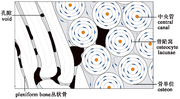
图1 骨组织基本结构
Fig.1 Basic structure of bone tissue
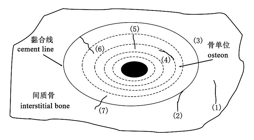
图2 骨单位中微裂纹的分类[41] 1.骨基质中的微裂纹 microcracks in the interstitial bone matrix;2.基质和黏合线之间的微裂纹 microcracks extending between the matrix and the cement line;3.沿黏合线延伸的微裂纹 microcracks along the cement line;4.层间微裂纹 interlamellar microcracks;5.接近但未到达黏合线的微裂纹 microcracks within the osteon approaching but not reaching the cement line;6.终止于黏合线的微裂纹 microcracks within the osteon terminating at the cement line;7.穿过黏合线的微裂纹 microcracks crossing the cement line.
Fig.2 Classification of micro-cracks in Haversian bone
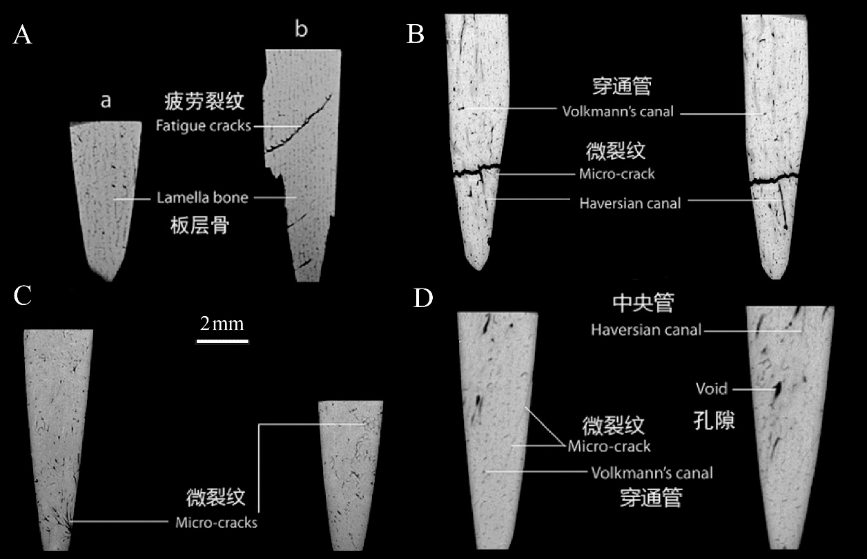
图3 CT扫描几种实验下微裂纹示意图[9] A.疲劳试验前后CT扫描图像,左侧(a)为未处理的对照组,右侧(b)为增加载荷直至骨骼断裂的实验组 CT scan images before and after fatigue testing: the left side (a) represents the untreated group, while the right side (b) shows the experimental group subjected to increasing load until bone fracture;B.兽皮穿刺实验 hide piercing experiment;C.模拟动物踩踏实验 trampling experiment;D.风化状态 weathering experiment。
Fig.3 CT scan images of microfractures from various experiments
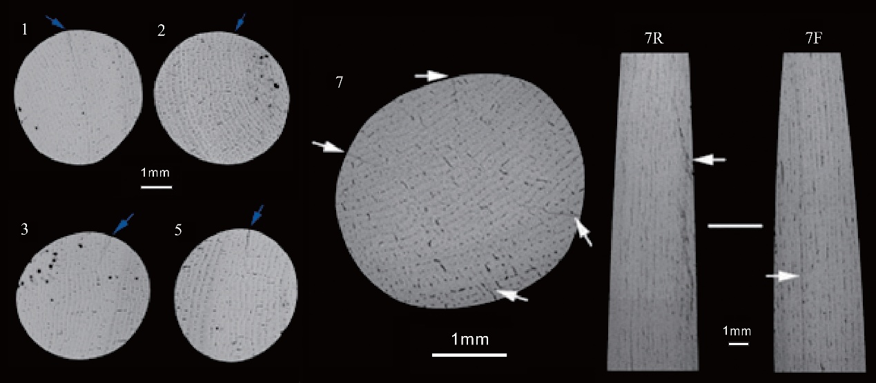
图4 狩猎实验(标本1,2,3,5)和加热实验后的骨箭头(标本7)内部显微结构[48] 箭头指向微裂纹的具体位置;数字表示实验过程中的箭头序号The arrows indicate the specific locations of microfractures, while the numbers correspond to the arrows used in the experimental process。F.前面front;R.右面right
Fig.4 Microfracture morphology of bone arrowheads after hunting experiments (specimens 1, 2, 3, 5) and heating experiments (specimen 7)
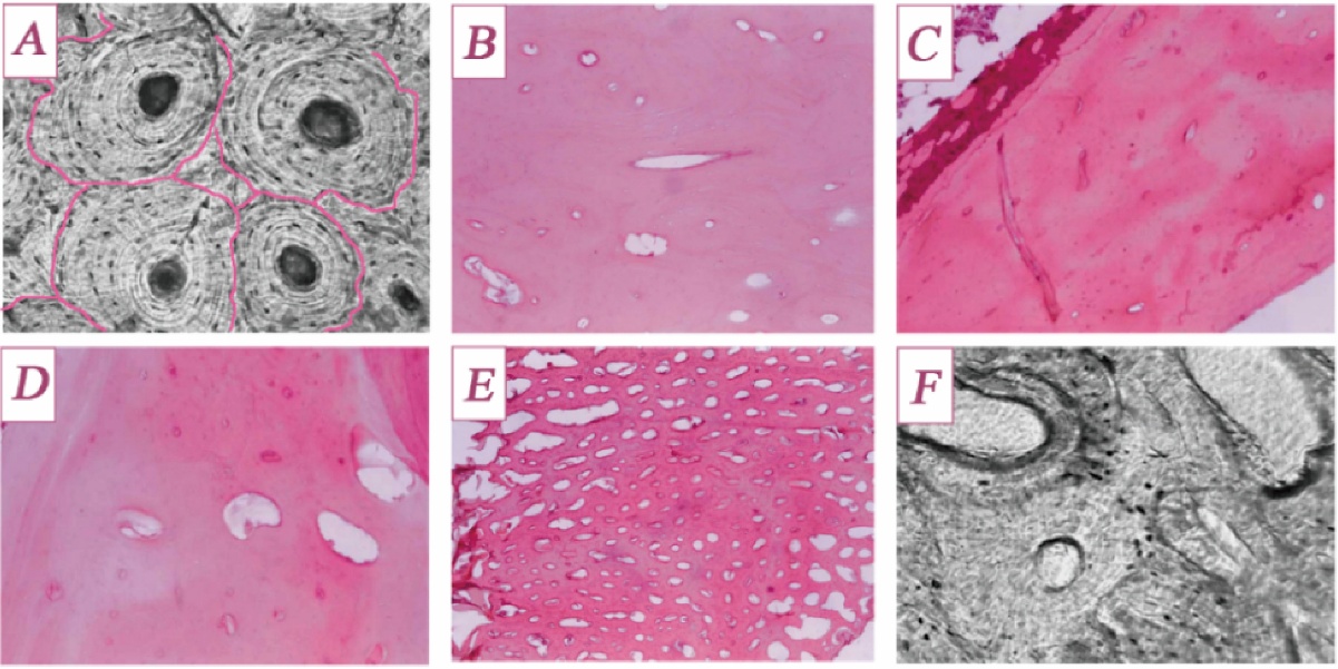
图5 比较人类和非人类长骨(胫骨)的组织学差异[69,71] A.人:高亮部分表示黏合线,标志着新旧骨单位的叠压关系 Human: The highlighted areas indicate cement lines, marking the overlapping relationship between new and old osteons;B.牛:骨单位形状近似椭圆形,中央管形状不规则 Cattle: Osteons are approximately oval-shaped, with irregularly shaped central canals;C.猫:单位面积骨单位分布密度高,中央管直径较小 Cat: High-density distribution of osteons per unit area, with smaller central canal diameters;D.狗:较高密度中等大小的骨单位 Dog: Moderately sized osteons with relatively high density;E.鸡:高密度分布的较小的骨单位 Chicken: Small osteons densely distributed;F.猪:骨单位形状不规则,周围有吸收腔隙 Pig: Irregularly shaped osteons with resorption cavities surrounding them.
Fig.5 Comparison of the histological differences between human and non-human bones
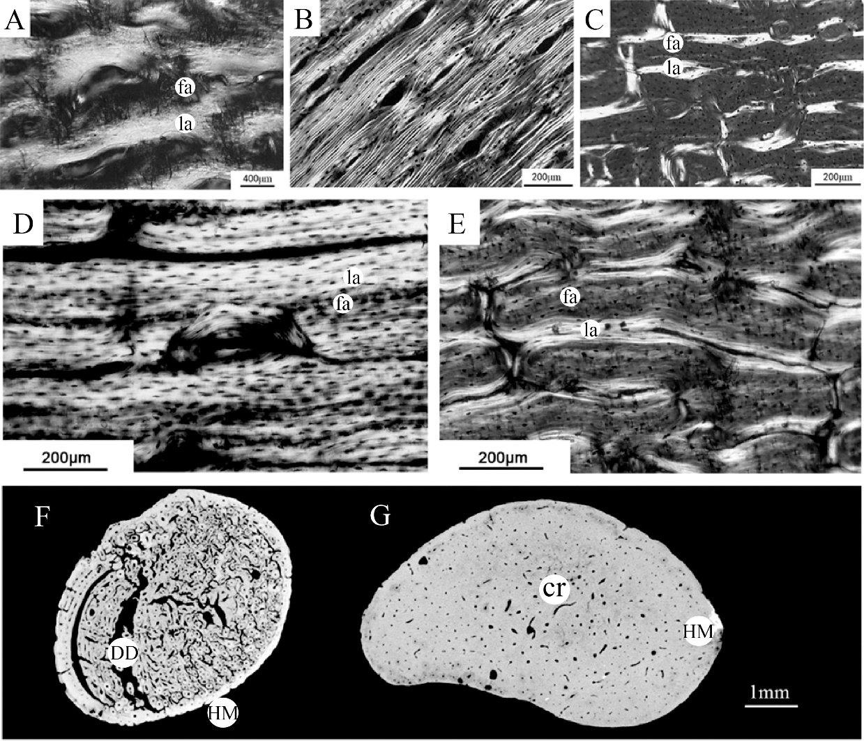
图6 不同个体的骨组织结构对比[78-80] A. 4天大婴儿股骨的骨组织结构 Bone tissue structure of a 4-day-old infant femur;B. 4岁半儿童肱骨的骨组织结构 Bone tissue structure of a 4.5-year-old child’s humerus;C.幼年猪桡骨的骨组织结构 Bone tissue structure of a juvenile pig’s radius;D.马骨中以层状成分为主的纤维板层骨 Fibrolamellar bone in horse bone, primarily composed of lamellar components;E.牛骨中以纤维成分为主的纤维板层骨 Fibrolamellar bone in cattle bone, primarily composed of fibrous components;F.偶蹄类 Artiodactyla;G.奇蹄类 Perissodactyla。fa.纤维成分 fibrous component;la.层状成分 lamellar component;cr.热裂纹 heat-induced cracking;HM.过度矿化 hyper-mineralisation;DD.成岩溶蚀作用 digenetic dissolution。
Fig.6 Comparison of bone tissue structures among different individuals
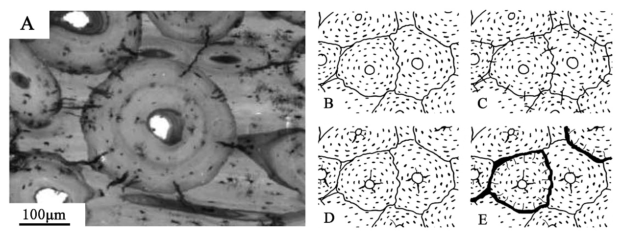
图7 骨骼化石在成岩过程中产生的几种微裂纹[91] A.早期成岩作用产生的径向微裂纹Radial microfractures caused by early diagenesis;B.新鲜状态下的哈弗氏骨Haversian bone in a fresh state;C. 在水生条件下,由于骨胶原蛋白吸水膨胀产生的微裂纹Under aquatic conditions radial microcracks appear due to water uptake and swelling of the bone collagen in secondary osteons;D.在干燥条件下,由于骨胶原蛋白收缩而产生的由中央管向外放射的微裂纹Under dry conditions shrinkage of the bone collagen in secondary osteons due to desiccation produces radial cracks from the Haversian canal outwards;E.干燥环境维持较长时间的情况下,周向裂纹将骨单位与周围分离,周缘出现微小的裂纹If desiccation is prolonged, circumferential cracks can isolate osteons from adjacent structures; small peripheral cracks occur, which start at the osteon boundary but do not spread beyond it
Fig.7 The types of microfractures occurring in bone fossils during the diagenetic process
| [1] | Gates St-Pierre C, Walker RB. Bones as Tools: Current Methods and Interpretations in Worked Bone Studies[J]. Oxford: BAR Publishing, 2007 |
| [2] | Backwell LR, d’Errico F. The first use of bone tools: a reappraisal of the evidence from Olduvai Gorge, Tanzania[J]. Palaeontologia africana, 2004, 40(9): 95-158 |
| [3] | Chomko SA. Bone “Awls” and utilized antler tines from Arnold research cave, 23Cy64, Missouri[J]. Plains Anthropologist, 1975, 20(67): 27-40 |
| [4] | LeMoine GM. Use wear on bone and antler tools from the Mackenzie Delta, Northwest Territories[J]. American Antiquity, 1994, 59(2): 316-334 |
| [5] | Bradfield J, Lombard M. A macrofracture study of bone points used in experimental hunting with reference to the South African Middle Stone Age[J]. South African Archaeological Bulletin, 2011, 66 (193): 67-76 |
| [6] | Wilkins J, Schoville BJ, Brown KS, et al. Evidence for early hafted hunting technology[J]. Science, 2012, 338 (6109): 942-946 |
| [7] | Forssman T. A macro-fracture investigation of the backed stone tools from Dzombo Shelter, eastern Botswana[J]. Journal of Archaeological Science: Reports, 2015, 3: 265-274 |
| [8] | 夏蒙棼, 韩闻生, 柯孚久, 等. 统计细观损伤力学和损伤演化诱致突变[J]. 力学进展, 1995, 1: 1-40 |
| [9] | Bradfield J. Investigating the potential of micro-focus computed tomography in the study of ancient bone tool function: results from actualistic experiments[J]. Journal of Archaeological Science, 2013, 40(6): 2606-2613 |
| [10] | Ritman EL. Micro-computed tomography—current status and developments[J]. Annu. Rev. Biomed. Eng, 2004, 6(1): 185-208 |
| [11] | Bera B, Mitra SK, Vick D. Understanding the micro structure of Berea Sandstone by the simultaneous use of micro-computed tomography (micro-CT) and focused ion beam-scanning electron microscopy (FIB-SEM)[J]. Micron, 2011, 42(5): 412-418 |
| [12] | Sutton MD. Tomographic techniques for the study of exceptionally preserved fossils[J]. Proceedings of the Royal Society B: Biological Sciences, 2008, 275 (1643): 1587-1593 |
| [13] | Bello SM, De Groote I, Delbarre G. Application of 3-dimensional microscopy and micro-CT scanning to the analysis of Magdalenian portable art on bone and antler[J]. Journal of Archaeological Science, 2013, 40(5): 2464-2476 |
| [14] | Lam YM, Chen X, Marean CW, et al. Bone density and long bone representation in archaeological faunas: comparing results from CT and photon densitometry[J]. Journal of Archaeological Science, 1998, 25(6): 559-570 |
| [15] | Beck L, Cuif JP, Pichon L, et al. Checking collagen preservation in archaeological bone by non-destructive studies (Micro-CT and IBA)[J]. Nuclear Instruments and Methods in Physics Research Section B: Beam Interactions with Materials and Atoms, 2012, 273: 203-207 |
| [16] | Villagran XS, Strauss A, Alves M, et al. Virtual micromorphology: The application of micro-CT scanning for the identification of termite mounds in archaeological sediments[J]. Journal of Archaeological Science: Reports, 2019, 24: 785-795 |
| [17] | Bradfield J, Wurz S. A functional assessment of the notched bone artefacts from Klasies River Main site[J]. South African Archaeological Bulletin, 2020, 75(213): 128-136 |
| [18] | Booth TJ, Redfern RC, Gowland RL. Immaculate conceptions: Micro-CT analysis of diagenesis in Romano-British infant skeletons[J]. Journal of Archaeological Science, 2016, 74: 124-134 |
| [19] | Currey JD. Bones: structure and mechanics[M]. Princeton university press, 2002 |
| [20] | Bell LS, Cox G, Sealy J. Determining isotopic life history trajectories using bone density fractionation and stable isotope measurements: a new approach[J]. American Journal of Physical Anthropology: The Official Publication of the American Association of Physical Anthropologists, 2001, 116(1): 66-79 |
| [21] | Sorg MH, Haglund WD. Forensic taphonomy: the postmortem fate of human remains[M]. Boca Raton: CRC Press, 1996 |
| [22] | Martin RB, Burr DB, Sharkey NA, et al. Skeletal tissue mechanics[M]. Springer, 1998, Vol 190 |
| [23] | Schultz M. Paleohistopathology of bone: a new approach to the study of ancient diseases[J]. American Journal of Physical Anthropology: The Official Publication of the American Association of Physical Anthropologists, 2001, 116(S33): 106-147 |
| [24] | Currey JD. The many adaptations of bone[J]. Journal of biomechanics, 2003, 36(10): 1487-1495 |
| [25] | Enlow DH. A comparative histological study of fossil and recent bone tissues[M]. Texas A&M University, 1955 |
| [26] | Enlow D. An evaluation of the use of bone histology in forensic medicine and anthropology[J]. Studies on the Anatomy and Function of Bone and Joints: Springer, 1966, 93-112 |
| [27] | Mulhern DM, Ubelaker DH. Differentiating human from nonhuman bone microstructure[J]. Bone histology: An anthropological perspective, 2012, 109-134 |
| [28] | Francillon-Vieillot H, de Buffrénil V, Castanet J, et al. Microstructure and mineralization of vertebrate skeletal tissues[J]. Skeletal biomineralization: patterns, processes and evolutionary trends, 1990, 1: 471-530 |
| [29] | Martin RB, Burr DB. Structure, function, and adaptation of compact bone[M]. New York: Raven Press, 1989 |
| [30] | Bell LS, Skinner MF, Jones SJ. The speed of post mortem change to the human skeleton and its taphonomic significance[J]. Forensic science international, 1996, 82(2): 129-140 |
| [31] | Jans MM, Nielsen-Marsh CM, Smith CI, et al. Characterisation of microbial attack on archaeological bone[J]. Journal of Archaeological Science, 2004, 31(1): 87-95 |
| [32] | Bell L, Boyde A, Jones S. Diagenetic alteration to teeth in situ illustrated by backscattered electron imaging[J]. Scanning, 1991, 13(2): 173-183 |
| [33] | Bradtmiller B, Buikstra JE. Effects of burning on human bone microstructure: a preliminary study[J]. Journal of Forensic Sciences, 1984, 29(2): 535-540 |
| [34] | Cattaneo C, DiMartino S, Scali S, et al. Determining the human origin of fragments of burnt bone: a comparative study of histological, immunological and DNA techniques[J]. Forensic science international, 1999, 102(2-3): 181-191 |
| [35] | Tersigni MA. Frozen human bone: A histological investigation[D]. Knoxville: University of Tennessee, 2002 |
| [36] | Andrews P, Evans EN. Small mammal bone accumulations produced by mammalian carnivores[J]. Paleobiology, 1983, 9(3): 289-307 |
| [37] | Andrews P. Owls, Caves and Fossils[M]. London: Natural History Museum Publications, 1990 |
| [38] | Kim JH, Niinomi M, Akahori T, et al. Effect of microstructure on fatigue strength of bovine compact bones[J]. JSME International Journal Series A Solid Mechanics and Material Engineering, 2005, 48(4): 472-480 |
| [39] | Currey JD. The structure and mechanics of bone[J]. Journal of Materials Science, 2012, 47: 41-54 |
| [40] | Li S, Demirci E, Silberschmidt VV. Variability and anisotropy of mechanical behavior of cortical bone in tension and compression[J]. Journal of the mechanical behavior of biomedical materials, 2013, 21: 109-120 |
| [41] | Norman TL, Wang Z. Microdamage of human cortical bone: incidence and morphology in long bones[J]. Bone, 1997, 20(4): 375-379 |
| [42] | Wheatley BP. Perimortem or postmortem bone fractures? An experimental study of fracture patterns in deer femora[J]. Journal of Forensic Sciences, 2008, 53(1): 69-72 |
| [43] | Lipson SF, Katz JL. The relationship between elastic properties and microstructure of bovine cortical bone[J]. Journal of biomechanics, 1984, 17(4): 231-240 |
| [44] | Lee TC, Mohsin S, Taylor D, et al. Detecting microdamage in bone[J]. Journal of anatomy, 2003, 203(2): 161-172 |
| [45] | 殷之平(著). 结构疲劳与断裂[M]. 西安: 西北工业大学出版社, 2012, 129 |
| [46] | Kasiri S, Taylor D. A critical distance study of stress concentrations in bone[J]. Journal of biomechanics, 2008, 41(3): 603-609 |
| [47] | Landrigan MD, Li J, Turnbull TL, et al. Contrast-enhanced micro-computed tomography of fatigue microdamage accumulation in human cortical bone[J]. Bone, 2011, 48(3): 443-450 |
| [48] | Backwell L, Bradfield J, Carlson KJ, et al. The antiquity of bow-and-arrow technology: evidence from Middle Stone Age layers at Sibudu Cave[J]. Antiquity, 2018, 92(362): 289-303 |
| [49] | Bradfield J, Lombard M, Reynard J, et al. Further evidence for bow hunting and its implications more than 60 000 years ago: Results of a use-trace analysis of the bone point from Klasies River Main site, South Africa[J]. Quaternary Science Reviews, 2020, 236: 106295 |
| [50] | Bradfield J, Hoffman J, De Beer F. Verifying the potential of micro-focus X-ray computed tomography in the study of ancient bone tool function[J]. Journal of Archaeological Science: Reports, 2016, 5: 80-84 |
| [51] | Pargeter J, Bradfield J. The effects of class I and II sized bovids on macrofracture formation and tool displacement: results of a trampling experiment in a southern African Stone Age context[J]. Journal of Field Archaeology, 2012, 37(3): 238-251 |
| [52] | Bradfield J, Brand T. Results of utilitarian and accidental breakage experiments on bone points[J]. Archaeological and Anthropological Sciences, 2015, 7: 27-38 |
| [53] | Behrensmeyer AK. Taphonomic and ecologic information from bone weathering[J]. Paleobiology, 1978, 4(2): 150-162 |
| [54] | Kenyeres B, Hegyi M. Unterscheidung des menschlichen und tierischen Knochengewebes[J]. Vierteljahresschr Gerichtsmed, 1903, 26: 225-232 |
| [55] | Harsányi L. Differential diagnosis of human and animal bone[A]. In: Grupe G, Garland AN. Histology of Ancient Human Bone: Methods and Diagnosis[C]. Palaeohistology Workshop, 3–5 October 1990, Göttingen. Berlin, Heidelberg: Springer, 1993, 79-94 |
| [56] | Dittmann K. Histomorphometrische Untersuchung der Knochenmikrostruktur von Primaten und Haustieren mit dem Ziel der Speziesidentifikaton unter Berücksichtigung von Domestikationseffekten[J]. Anthropologischer Anzeiger, 2003, 175-188 |
| [57] | Urbanová P, Novotný V. Distinguishing between human and non-human bones: histometric method for forensic anthropology[J]. Anthropologie, 2005, 43(1): 77-86 |
| [58] | Sauer NJ, Lackey WL. Anthropology:skeletal analysis[A]. In: Siegel JA, Saukko PJ, Knupfer GC(Eds.). Encyclopedia of Forensic Sciences[M]. New York: Academic Press, 2000: 261-270 |
| [59] | Owsley DW, Mires AM, Keith M. Case involving differentiation of deer and human bone fragments[J]. Journal of Forensic Sciences, 1985, 30(2): 572-578 |
| [60] | Mulhern DM, Ubelaker DH. Differences in osteon banding between human and nonhuman bone[J]. Journal of Forensic Sciences, 2001, 46(2): 220-222 |
| [61] | Andronowski JM, Pratt IV, Cooper DM. Occurrence of osteon banding in adult human cortical bone[J]. American journal of physical anthropology, 2017, 164(3): 635-642 |
| [62] | Demeter G, Mátyás J. Mikroskopisch vergleichend-anatomische Studien an Röhrenknochen mit besonderer Rücksicht auf die Unterscheidung menschlicher und tierischer Knochen[J]. Zeitschrift für Anatomie und Entwicklungsgeschichte, 1928, 87(1): 45-99 |
| [63] | Jowsey J. Studies of Haversian systems in man and some animals[J]. Journal of anatomy, 1966, 100(Pt 4): 857 |
| [64] | Singh I, Tonna E, Gandel C. A comparative histological study of mammalian bone[J]. Journal of Morphology, 1974, 144(4): 421-437 |
| [65] | Martiniakova M, Grosskopf B, Omelka R, et al. Histological study of compact bone tissue in some mammals: a method for species determination[J]. International Journal of Osteoarchaeology, 2007, 17(1): 82-90 |
| [66] | Dominguez VM, Crowder CM. The utility of osteon shape and circularity for differentiating human and non-human Haversian bone[J]. American journal of physical anthropology, 2012, 149(1): 84-91 |
| [67] | Martiniakova M, Grosskopf B, Omelka R, et al. Differences among species in compact bone tissue microstructure of mammalian skeleton: use of a discriminant function analysis for species identification[J]. Journal of Forensic Sciences, 2006, 51(6): 1235-1239 |
| [68] | Crescimanno A, Stout SD. Differentiating fragmented human and nonhuman long bone using osteon circularity[J]. Journal of Forensic Sciences, 2012, 57(2): 287-294 |
| [69] | Hillier ML, Bell LS. Differentiating human bone from animal bone: a review of histological methods[J]. Journal of Forensic Sciences, 2007, 52(2): 249-263 |
| [70] | Stan E, Muresan CO, Daescu E, et al. A Review of Histological Techniques for Differentiating Human Bone from Animal Bone[J]. Methods and Protocols, 2024, 7(4): 51 |
| [71] | Morales JP, Roa H, Zavando D, et al. Determination of the species from skeletal remains through histomorphometric evaluation and discriminant analysis[J]. Int J Morphol, 2012, 30(3): 1035-1041 |
| [72] | Pfeiffer S, Pinto D. Histological analyses of human bone from archaeological contexts[A]. In: Stout SD(Ed). Bone Histology[M]. Boca Raton: CRC Press, 2011: 343-364 |
| [73] | Halstead P, Collins P, Isaakidou V. Sorting the sheep from the goats: Morphological distinctions between the mandibles and mandibular teeth of AdultOvis and Capra[J]. Journal of Archaeological Science, 2002, 29(5): 545-553 |
| [74] | Zedda M, Brits D, Giua S, et al. Distinguishing domestic pig femora and tibiae from wild boar through microscopic analyses[J]. Zoomorphology, 2019, 138(1): 159-170 |
| [75] | Hollund HI, Jans MM, Collins MJ, et al. What happened here? Bone histology as a tool in decoding the postmortem histories of archaeological bone from Castricum, The Netherlands[J]. International Journal of Osteoarchaeology, 2012, 22(5): 537-548 |
| [76] | Cuijpers A. Histological identification of bone fragments in archaeology: telling humans apart from horses and cattle[J]. International Journal of Osteoarchaeology, 2006, 16(6): 465-480 |
| [77] | Burke A, Drapeau MS. The Histology of Skeletal Tissues as a Tool in Paleoanthropological and Archaeological Investigations[J]. Vertebrate Skeletal Histology and Paleohistology: CRC Press, 2021, 781-792 |
| [78] | Cuijpers S. Distinguishing between the bone fragments of medium-sized mammals and children. A histological identification method for archaeology[J]. Anthropologischer Anzeiger, 2009, 181-203 |
| [79] | Cuijpers S, Lauwerier RC. Differentiating between bone fragments from horses and cattle: a histological identification method for archaeology[J]. Environmental Archaeology, 2008, 13(2): 165-179 |
| [80] | Bradfield J. Identifying animal taxa used to manufacture bone tools during the Middle Stone Age at Sibudu, South Africa: Results of a CT-rendered histological analysis[J]. PLoS One, 2018, 13(11): e0208319 |
| [81] | Guan Y, Gao X, Li F, et al. Modern human behaviors during the late stage of the MIS3 and the broad spectrum revolution: evidence from a Shuidonggou Late Paleolithic site[J]. Chinese Science Bulletin, 2012, 57: 379-386 |
| [82] | Song Y, Li X, Wu X, et al. Bone needle fragment in LGM from the Shizitan site (China): Archaeological evidence and experimental study[J]. Quaternary International, 2016, 400: 140-148 |
| [83] | Zhang S, d'Errico F, Backwell LR, et al. Ma'anshan cave and the origin of bone tool technology in China[J]. Journal of Archaeological Science, 2016, 65: 57-69 |
| [84] | Zhang S, Doyon L, Zhang Y, et al. Innovation in bone technology and artefact types in the late Upper Palaeolithic of China: Insights from Shuidonggou Locality 12[J]. Journal of Archaeological Science, 2018, 93: 82-93 |
| [85] | Orlando L, Allaby R, Skoglund P, et al. Ancient DNA analysis[J]. Nature reviews methods primers, 2021, 1(1): 14 |
| [86] | Richter KK, Codlin MC, Seabrook M, et al. A primer for ZooMS applications in archaeology[J]. Proceedings of the National Academy of Sciences, 2022, 119(20): e2109323119 |
| [87] | Jacobson L, De Beer FC, Nshimirimana R, et al. Neutron tomographic assessment of incisions on prehistoric stone slabs: a case study from Wonderwerk Cave, South Africa[J]. Archaeometry, 2013, 55(1): 1-13 |
| [88] | Caruso V, Marinoni N, Diella V, et al. Bone diagenesis in archaeological and contemporary human remains: an investigation of bone 3D microstructure and minero-chemical assessment[J]. Archaeological and Anthropological Sciences, 2020, 12: 1-18 |
| [89] | Keenan SW. From bone to fossil: A review of the diagenesis of bioapatite[J]. American Mineralogist, 2016, 101(9): 1943-1951 |
| [90] | Pfretzschner HU. Fossilization of Haversian bone in aquatic environments[J]. Comptes Rendus Palevol, 2004, 3(6-7): 605-616 |
| [91] | Pfretzschner HU, Tütken T. Rolling bones-Taphonomy of Jurassic dinosaur bones inferred from diagenetic microcracks and mineral infillings[J]. Palaeogeography, Palaeoclimatology, Palaeoecology, 2011, 310(1-2): 117-123 |
| [1] | 何湘栋, 梁越, 王春雪, 牛东伟, 杜雨薇. 蔚县盆地东沟遗址出土动物化石的初步研究[J]. 人类学学报, 2026, 45(03): 586-599. |
| [2] | 惠家明. 颅骨板障静脉的三维复原及其在人类演化中的意义[J]. 人类学学报, 2025, 44(04): 545-555. |
| [3] | 李璇, 顾雪军. 2015~2018年河南栾川龙泉洞遗址发掘简报[J]. 人类学学报, 2025, 44(04): 714-726. |
| [4] | 曹雨昕, 孙璐, 张乐, 张双权. 骨骼表面人类齿痕特征的实验研究[J]. 人类学学报, 2025, 44(02): 242-254. |
| [5] | 王颖, 张乐, 杨石霞, 张双权. 山西峙峪遗址出土动物骨骼的埋藏学[J]. 人类学学报, 2025, 44(02): 255-269. |
| [6] | 张乐, 张双权. 贵州普定穿洞遗址1981年出土的骨制品[J]. 人类学学报, 2024, 43(06): 1048-1063. |
| [7] | 韩芳, 李冀源, 乔虹, 徐海伦, 何虹霖, 高璇, 吕红亮, 杜战伟, 蔡林海, 甄强, 马文灵. 环青海湖地区细石叶遗存新发现[J]. 人类学学报, 2024, 43(05): 839-852. |
| [8] | 王华, 李占扬, Thijs van KOLFSCHOTEN. 德国西宁根与中国灵井的骨器比较[J]. 人类学学报, 2024, 43(02): 214-232. |
| [9] | 杜雨薇, 张乐, 叶芷, 裴树文. 蔚县盆地吉家庄旧石器遗址动物骨骼的埋藏学分析[J]. 人类学学报, 2023, 42(03): 359-372. |
| [10] | 张乐, 吴秀杰, 张双权. 四川资阳人遗址出土的骨锥[J]. 人类学学报, 2023, 42(01): 1-14. |
| [11] | 梁琪瑶, 张伟, 陈全家, 田禾. 黑龙江齐齐哈尔洪河遗址出土的骨器[J]. 人类学学报, 2021, 40(05): 751-763. |
| [12] | 戴静雯, 张双权, 张乐. 史前人类对动物骨骼油脂的开发和利用[J]. 人类学学报, 2021, 40(03): 503-512. |
| [13] | 黄超, 张双权. X射线衍射技术在烧骨实验研究中的初步应用[J]. 人类学学报, 2021, 40(03): 513-525. |
| [14] | 潘雷, 廖卫, 王伟, 刘建辉, 吉学平, 杨晓梅, 郝以鑫. 禄丰古猿蝴蝶种下第四前臼齿釉质-齿质交界面的三维几何形态[J]. 人类学学报, 2020, 39(04): 555-563. |
| [15] | Vijaya Lakshmi Pavani MOLLI, Anubhav JAIN, 傅江南, 吴英杰, Jian Q.FENG, 王谦. 树鼩的骨元和骨细胞穴微观结构超微影像的定性比较[J]. 人类学学报, 2020, 39(04): 564-575. |
| 阅读次数 | ||||||
|
全文 |
|
|||||
|
摘要 |
|
|||||
 京ICP证05002819号-3